Cervical Spondylosis (Arthritis of the Neck)

Neck pain is extremely common. It can be caused by many things, and is most often related to getting older.

Like the rest of the body, the bones in the neck (cervical spine) slowly degenerate as we age. This frequently results in arthritis. Arthritis of the neck is called cervical spondylosis.

Cervical spondylosis is the degeneration of the joints in the neck. It becomes increasingly more common as people age. More than 85% of people over age 60 are affected.

Although it is a form of arthritis, cervical spondylosis rarely becomes a crippling or disabling type.

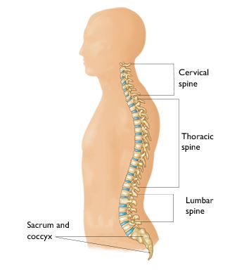
Understanding your spine and how it works can help you better understand cervical spondylosis.

Cause

Disk Degeneration and Bone Spurs


In the spine, arthritis can result as the disk degenerates and loses water content. In children and young adults, disks have high water content. As we get older, our disks begin to dry out and weaken. This problem causes settling, or collapse, of the disk spaces and loss of disk space height.

As the facet joints experience increased pressure, they also begin to degenerate and develop arthritis, similar to the hip or knee joint. The cartilage that covers and protects the joints wears away.

If the cartilage wears away completely, it can result in bone rubbing on bone. To make up for the lost cartilage, your body may respond by growing new bone in your facet joints to help support the vertebrae. Over time, this bone overgrowth — called spurs — may narrow the space for the nerves to pass through (stenosis).


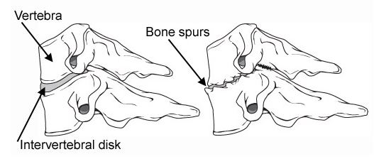
(Left) Side view of a healthy cervical vertebra and disk. (Right) A disk that has degenerated and collapsed.

Risk Factors

There are several factors that increase your risk for cervical spondylosis. The following have all been linked to higher risks of neck pain and spondylosis:

  • Genetics – if your family has a history of neck pain
  • Smoking – clearly linked to increased neck pain
  • Occupation – jobs with lots of neck motion and overhead work
  • Mental health issues – depression/anxiety
  • Injuries/trauma – car wreck or on-the-job injury

Symptoms

Pain from cervical spondylosis can be mild to severe. It is sometimes worsened by looking up or down for a long time, or with activities such as driving or reading a book. It also feels better with rest or lying down.

Additional symptoms include:

  • Neck pain and stiffness (may be worse with activity)
  • Numbness and weakness in arms, hands, and fingers
  • Trouble walking, loss of balance, or weakness in hands or legs
  • Muscle spasms in neck and shoulders
  • Headaches
  • Grinding and popping sound/feeling in neck with movement

Doctor Examination

Determining the source of the pain is essential to recommend the appropriate treatment and rehabilitation. Therefore, a comprehensive examination is required to determine the cause of neck pain.

Your doctor will take a complete history of the difficulties you are having with your neck. He or she may ask you about other illnesses or injuries that occurred to your neck. Questions may include: When did your neck begin to hurt? Has it ever hurt like this before? When your neck hurts, how often and for how long does it hurt? Does anything make it better or worse? Were you ever involved in an accident or had an injury to your neck? Have you ever been treated for your neck pain?

A thorough physical exam will include your neck, shoulders, arms, and frequently your legs, as well. Your strength, touch sensation, reflexes, blood flow, flexibility of your neck and arms as well as your walking may be tested. The doctor may press on your neck and shoulders, and feel for trigger (tender) points or swollen glands.

Tests

Your doctor may supplement your evaluation with blood tests, and, if necessary, consult with other medical specialists. Other tests which may help your doctor confirm your diagnosis include:

X-rays

These pictures are traditionally ordered as a first step in imaging the spine. X-rays will show aging changes, like loss of disk height or bone spurs.

Magnetic resonance imaging (MRI)

This study can create better images of soft tissues, such as muscles, disks, nerves, and the spinal cord.

Computed tomography (CT) scans

This specialized x-ray study allows careful evaluation of the bone and spinal canal.

Myelography

This specific x-ray study involves injecting dye or contrast material into the spinal canal. It allows for careful evaluation of the spinal canal and nerve roots.

Electromyography (EMG)

Nerve conduction studies and electromyography may be performed by another doctor to look for nerve damage or pinching.

Treatment

Nonsurgical Treatment

Physical therapy. Strengthening and stretching weakened or strained muscles is usually the first treatment that is advised. Your physical therapist may also use cervical (neck) traction and posture therapy. Physical therapy programs vary, but they generally last from 6 to 8 weeks. Sessions are scheduled 2 to 3 times a week.

Medications. Several medications may be used together during the first phase of treatment to address both pain and inflammation.

  • Acetaminophen. Mild pain is often relieved with acetaminophen.
  • Non-steroidal anti-inflammatory drugs (NSAIDs). Often prescribed with acetaminophen, drugs like ibuprofen and and naproxen are considered first-line medicines for neck pain. They address both pain and swelling, and may be prescribed for a number of weeks, depending on the specific problem. Other types of pain medicines can be considered if you have serious contraindications to NSAIDs, or your pain is not well controlled.
  • Muscle relaxants. Medications such as cyclobenzaprine or carisoprodol can also be used in the case of painful muscle spasms.

Soft Collars. These collars limit neck motion and allow the muscles of the neck to rest. Soft collars should only be worn for short periods of time because long-term wear can decrease the strength of neck muscles.

Ice, heat, other modalities. Careful use of ice, heat, massage, and other local therapies can help relieve symptoms.

Steroid-Based Injections. Many patients find short-term pain relief from steroid injections. Various types of these injections are routinely performed. The most common procedures for neck pain include:

Cervical epidural block. In this procedure, steroid and anesthetic medicine is injected into the space next to the covering of the spinal cord (“epidural” space). This procedure is typically used for neck and/ or arm pain that may be due to a cervical disk herniation, also known as radiculopathy or a “pinched nerve.”

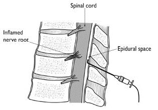
Epidural injection in the cervical spine.

Cervical facet joint block. Steroid and anesthetic medicine is injected into the capsule of the facet joint in this procedure. The facet joints are located in the back of the neck and provide stability and movement. These joints can develop arthritic changes that may contribute to neck pain.

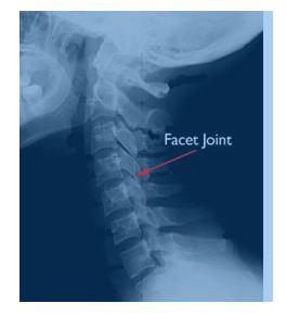
Cervical facet joint.

Medial branch block and radiofrequency ablation. This procedure is used in some cases of chronic neck pain. It can be used for both diagnosis and treatment of a potentially painful joint.

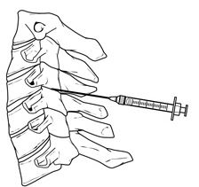
Facet joint injection in the cervical spine.

During the diagnosis portion of the procedure, the nerve that supplies the facet joint is blocked with a local anesthetic (like the medicine used by your dentist). Your doctor will ask if your neck pain is completely gone. If so, then your doctor has pin-pointed the source of your neck pain. The next step is to block the pain more permanently. This is done by damaging the nerve with radiofrequency, a procedure called radiofrequency ablation.

Although less invasive than surgery, steroid-based injections are prescribed only after a complete evaluation by your doctor. Before considering these injections, discuss with your doctor the risks and benefits of these procedures for your specific condition.

Surgical Treatment

It is uncommon for people with only cervical spondylosis and neck pain to be treated with surgery. Surgery is reserved for patients who have severe pain that has not been relieved by other treatment. Some patients with severe pain will unfortunately not be candidates for surgery. This is due to the widespread nature of their arthritis, other medical problems, or other causes for their pain, such as fibromyalgia.

People who have progressive neurologic symptoms, such as weakness, numbness, or falling, are more likely to be helped by surgery.

Reference: American Academy of Orthopaedic Surgeons. http://orthoinfo.aaos.org/topic.cfm?topic=A00369

Lumbar Spondylosis without Myelopathy

Lumbar spondylosis is broadly defined as osteoarthritic changes affecting the triad of joints forming the spinal columns: the paired zygapophysial joints (z-joints) posteriorly and the intervertebral disk anteriorly. These degenerative changes are ubiquitous with increasing age, but in some cases they can be associated with low back pain (LBP). This condition can cause secondary myelopathy if the spondylosis results in cauda equina compression.

Etiology

The exact cause of lumbar spondylosis is unknown. Because it affects synovial joints, lumbar spondylosis can be thought of as being analogous to peripheral osteoarthritis (OA), and likely results from an imbalance in the synthesis and degeneration of the articular cartilage.

Epidemiology including risk factors and primary prevention

The prevalence of radiographic spondylosis increases with age. It is uncommon in the first few decades of life, but ubiquitous by the age of 65. In those with LBP, the prevalence ranges from 7% to 75%, depending on the diagnostic criteria. The prevalence in the United States ranges from 15% to 45% among patients with chronic LBP.

Risk Factors

Commonly accepted risk factors are the following: increasing age, genetic predisposition, being a woman, previous injury, and joint overload from malalignment and/or abnormal z-joint orientation. After the age of 45 years, advanced cartilage changes, subchondral sclerosis, and osteophyte formation are common. Genetic predisposition has also been shown to be a significant contribution to symptomatic and radiographic lumbar spondylosis in several studies. Cadaveric studies demonstrated that when compared with men, women of similar age showed a larger grade of cartilage degeneration and more osteophytes. The presence of estrogen receptors in cartilage was found, and higher estrogen expression correlates directly with higher levels of z-joint arthritis. Disk desiccation and z-joint malalignment are both associated with spondylosis. Greater z-joint sagittal orientation correlated more with degenerative changes in the z-joints and a higher incidence of spondylolisthesis.

Patho-anatomy/physiology

Studies have shown that within the lumbar spine this degradation frequently follows a common degenrative cascade, with the initial step being intervertebral disk desiccation. Collagen cross-linking and inability to hold water stiffens the cartilage and the capsule structures, leading to an altered range of motion. In the lumbar spine, the z-joints have a posterior-lateral orientation to resist axial rotation and translation. Thus, the degenerative changes frequently arise earlier and are more advanced in the regions that resist translation, specifically the anterior-medial portion of the joints and most frequently at the L4/L5 and L5/S1 levels, likely because of their proximity to a fused sacrum.

Disease progression including natural history, disease phases or stages, disease trajectory (clinical features and presentation over time)

Although variable, most of the patients experience a benign clinical course. Studies have shown the following:

  • Radiologic spondylosis directly increases with age, irrespective of pain.
  • Spondylosis was shown to be equally present in those with and without LBP.
  • No correlation between the magnitude of z-joint arthorpathy and the severity of pain.

Specific secondary or associated conditions and complications

z-joint hypertrophy, because of osteophyte formation combined with ligament redundancy from the disk desiccation, may collectively lead to spinal canal and/or intervertebral foramen narrowing. The increase in translation forces from disk desiccation, combined with the z-joints decreased ability to resist these factors, may also result in spinal instabilities, specifically spondylolisthesis. Excess synovial fluid production can result in secondary synovial cysts formation. All of these changes can result in central or lateral stenosis, radiculopathy, and/or, in severe cases, cauda equina syndrome.

Assessment

History

The symptoms are acute or gradual onset of diffuse LBP, possibly with an epicenter. Pain may refer unilaterally or bilaterally to the buttock, hip, groin, and thigh regions; although, typically, it does not extend past the knee. The pain tends to worsen with extension, rotation, and standing; it is better with lying and L-spine flexion. By definition, lumbar spondylosis exhibits no neurologic deficits; however, because of its association with conditions that can affect the neurologic function of the lower limbs, it is imperative to ask about weakness, balance, gait, and bowel/bladder function.

Physical examination

A systematic review revealed that most physical exam maneuvers have limited or no diagnostic validity for spondylosis. Paraspinal tenderness is the only physical exam maneuver that seems to correlate with z-joint arthropathy. Although clasically felt to diagnose z-joint pain, joint loading with pain on extension and ipsilateral rotation has been not shown to consistently correlate with spondylosis. Because the pain distribution may overlap with other clinical entities, a comprehensive exam including hip, sacroiliac joint provocative maneuvers should be performed routinely. Associated neurologic conditions should be ruled out through thorough strength, sensation, reflexes, gait, and balance testing.

Laboratory studies

None are routinely indicated. Laboratory studies, such as C-reactive protein, sedimentation rate, and complete blood count, can be done if there is a clinical suspicion for a more nefarious condition, such as tumor, infection, or rheumatologic disease, such as spondyloarthropathies.

Imaging

z-joint arthropathy is common on all imaging modalities with increasing age, and has a weak to no association with controlled diagnostic blocks. Imaging is therefore typically done to rule out other disorders, and can take the form of plain radiographs, magnetic resonance imaging (MRI), and computed tomorgraphy (CT) studies.

Plain radiographs are not sensitive for detecting early z-joint OA, but are highly accessible, and often used. Lateral views are important to study sagittal alignment and the presence of spondylolisthesis.

MRI is generally not needed for diagnosis of spondylosis, but it is useful for evaluating the soft tissues and neural elements within the spine. Large amounts of fluid within the z-joints may represent instability, which can be further assessed by flexion/extension films.

CT is typically utilized only when an MRI is unobtainable.

Supplemental assessment tools

Diagnostic Injections

Given the high prevalence of radiographic spondylosis, the only means for an accurate diagnosis of symptomatic lumbar spondylosis are controlled diagnostic blocks11 of the medial branch nerves that innervate the z-joints.

Several key principles exist for diagnostic blocks:

  • Because of inaccuracy of landmark guidance, all blocks should be preformed utilizing image guidance.
  • False positive blocks occur at a high rate of 17% to 41% in the lumbar spine, thus necessitating a second control block for confirmation of the diagnosis. The 2 injections are done with anesthetics with differing durations of effect. With dual positive blocks (>1h with lidocaine and >3h with bupivacaine hydrochloride), the sensitivity and specificity are greatly enhanced.
  • The greater the percentage of pain relief a patient obtains with a given injection, the more likely that injection correctly targeted the pain generator. Some clinicians accept 50% pain relief as a diagnostic criterion. An 80% pain relief with comparative medial branch blocks results in more successful outcomes with radiofrequency ablation.

Rehabilitation Management and Treatments

Available or current treatment guidelines

Although a few guidelines have been created, to date, none are universally accepted for z-joint treatment.

Coordination of care

As with all spine conditions, treatment should ideally be a multidisciplinary team and a coordinated approach with a physical therapist and a phsyician.

Patient & family education

Patients should be educated that the condition typically responds to treatment. Education should be provided regarding warning signs for disease progression into secondary neurologic conditions, such as myleopathy.

Emerging/unique Interventions

There are several validated outcome measures that can be utilized for grading functional limitations including the following: McGill Low Back Pain Scale, Oswestry Disability Index, and the Medical Outcomes Study 36-Item Short-Form Health Survey. These are typically administered at every office visit for following the functional limitations and disease progression.

Translation into practice: practice “pearls”/performance improvement in practice (PIPs)/changes in clinical practice behaviors and skills

Physicians should have a low threshold for ruling out other serious conditions that also commonly present as LBP in this age group. If injections are required and successful because of a failure of other treatments, patients should be encouragd to engage in daily therapuetic spine exercises to maximize function and hopefully prevent relapse of pain.

Cutting edge/emerging and unique concepts and practice

Cutting edge concepts and practice

New techniques and methods to enhance RFA lesion size while preserving safety are being developed. Limited literature exists on their efficacy, but 2 such technolgies are water-cooled RFA and Nimbus needles. These will hopefully enhance success rates and minimize technical failures. Studies are also currently underway on biologics to alter disease progression.

Gaps in the evidence-based knowledge

Given the limitations of imaging, physical exam, and prognostic blocks, individual characteristics that may help predict a successful response to a particular treatment are crucial. Also, more studies need to be done on those with block proven spondylosis, especially the conservative and interventional therapies.

Post-Laminectomy Syndrome

Post-laminectomy syndrome is a condition where the patient suffers from persistent pain in the back following surgery to the back. This article reviews this condition in a bit more detail.

A laminectomy is a procedure where a part of the vertebra that protects the spinal-cord is removed. It is usually performed to relieve pressure on the spinal-cord from a protruding disc. Very often, following a laminectomy, patients recover without any complications. However in a small group of people, back pain and sometimes leg pain may persist following laminectomy. This persistent pain is called post laminectomy syndrome. Post-laminectomy syndrome is also called Failed Back Surgery Syndrome, or FBSS.

What causes Post-laminectomy Syndrome?

There are a number of reasons why patients can develop back pain following a laminectomy. These could include the fact that the surgery itself was probably not necessary or that the surgery did not have the outcome that was expected. In some cases, despite performing a laminectomy, patients experience pain because the spinal column itself is narrowed in a condition called spinal stenosis. Sometimes, there may be a small fragment of the disc still remaining following the laminectomy which can irritate the spinal-cord causing pain. In other words, the term “post laminectomy syndrome” encompasses any cause that results in back pain following back surgery.

There is also evidence to suggest that patients who are smokers are more likely to develop post-laminectomy syndrome following surgery to the back. In addition to this, patients who have previously had a failed surgery in the back are at a higher risk of developing this syndrome.

Symptoms and Diagnosis

The most common symptom that patients experience is back pain at the site of surgery along with leg pain. As a result of the pain, patients have difficulty performing their activities of daily living and may have difficulty sleeping as well. The longer the pain lasts, the more of an impact it can have on the patient’s lives resulting in depression and anxiety.

When examined, patients will still complain of tenderness at the site of surgery. Doctors may notice altered postures and varied positions when walking.

In most cases, a history and clinical examination is sufficient to make a preliminary diagnosis. However, sometimes x-rays of the site of pain may reveal the cause for the pain. More advanced scan such as a CT scan or MRI will show whether there is residual compression on the spinal nerves following surgery. Any infections or abscess formation around the spinal cord may also be diagnosed this way though additional blood tests may be required.

How is Post-laminectomy Syndrome treated?

Treatment options may vary. Below are some of the options your doctor may consider.

  • Pain killers:these may be the first-line treatment that is prescribed by the doctors. In cases of post-laminectomy syndrome, the doctors may prescribe morphine-based painkillers in high doses. However, if this is done, the patient will be monitored closely to avoid the development of dependence on opioids.

  • Physical therapy:this may be recommended by the treating physician as physical therapists can prescribe exercises and treatments such as electrical stimulation which can help relieve pain and improve overall movement.

  • Specialist therapies: in some cases, doctors may consider referring patients for more advanced treatments such as epidural nerve blocks, radiofrequency denervation and platelet rich plasma therapy. Research is always being conducted with newer techniques being looked into all the time.


Lumbar Epidural Injection

What is an epidural space?

It is a space that surrounds the spinal cord and nerve roots. Furthermore, the back spine is separated into four regions: cervical (neck), thoracic (mid back), lumbar (low back), and caudal (sacral area). Therefore, the lumbar epidural injection defines the epidural space in the low back region.

What are the indications of lumbar epidural injection?

Among many indications the listed are the main indications: herniated disc, degenerated disc disease (DDD), radiculopathy, bone spurs, thicken ligamentum flavum, low back fusion, failed back surgery syndrome, chronic regional pain syndrome (RSD), diabetic neuropathy and spinal stenosis.

How does lumbar epidural steroid injection bring pain relief?

The pain is produced due to inflammation of the spinal nerve roots, which may be experienced as pins/needle sensation, shooting /stabbing pain which radiate from low back down to the legs and/or feet. With the lumbar epidural injection, steroids surround the lumbar spinal nerves, which help decrease the spinal nerve root inflammation and irritation. Therefore, the lumbar epidural injection can help decrease the low back and leg pain symptoms.

What are risks for the lumbar epidural injection?

Among many, here are few listed: increased pain, infection, bleeding, nerve damage, weakness.

How is the lumbar epidural injection performed?

After sterile preparation of the lumbar region, the injection site if localized under X-ray. Following the local anesthetic applied to the injection site, which can help decrease the injection site pain, the needle is guided toward the epidural space with the help of X-ray. After the space is localized, it can be further confirmed with liquid contrast, which can help further confirm the epidural space. After the space is confirmed, the steroid medication is injected, and the needle is taken out at the end of the procedure.

What to expect after the procedure?

This is an outpatient procedure. Patients are monitored in the recovery room after the procedure. Patient is educated that pain relief may be achieved to its full potential over the 1-2 week period. Patient may experience some localized mid-back pain at the injection site, which may be minimized by the previously applied local anesthetic. Patients are discharged from the recovery room once they have the discharge criteria.

How long the relief from the lumbar epidural steroid injection would last for?

It varies from patient to patient. Usually, the pain relief can last for 6-8 weeks, and may require repeat the epidural injections if necessary. Please tell your pain physician if you are taking any blood thinners. For example: Coumadin, Plavix, Heparin, Lovenox and etc. The blood thinners need to be stopped before the epidural injection, and the time frame will be prescribed by your pain physician.

Displacement of Cervical Disc Without Myelopathy

Displacement, Cervical Intervertebral Disc Without Myelopathy

Displacement of a cervical intervertebral disc refers to protrusion or herniation of the disc between two adjacent bones (vertebrae) of the cervical spine in the neck (vertebrae C2 through C7). Note that there is no disc between the skull and C1 or between C1 and C2. Although displacement is commonly referred to as a slipped disc, the disc does not actually slip.

The discs between each vertebra form a cushion that absorbs shock and allows movement of the neck. The discs are composed of an inner gel-like material (nucleus pulposus) and an outer ring of tough, fibrous material (annulus fibrosis). Sometimes the fibrous material develops a weak area that allows the nucleus pulposus to intrude into the spinal canal (disc displacement or herniation). Depending on the site of the intrusion, the disc may compress either the spinal cord or the exiting nerves, or both. Pressure on an exiting cervical nerve root where it exits the spinal canal can cause changes in sensory (touch, pinprick, temperature), motor (muscle strength), and reflex function in the innervated areas (upper limb). These types of changes are collectively referred to as radiculopathy; however, disc displacement may also occur without radiculopathy. Cervical radiculopathy also may be caused by tumors, infection, or vertebral fracture. Disruption of the annulus fibrosis itself may also cause symptoms (annular disruption, distension, or tear). This can allow the nucleus pulposus to leak out of the disc, causing an intense and painful chemical inflammation (radiculitis).

Disc herniations (commonly called “soft discs” in the neck) tend to occur in younger adults who have only mild loss of disc height (disc degeneration) and thus have enough disc material still present to produce a protrusion or herniation. The same radiculopathy symptoms and signs on exam can be produced without a disc herniation in older individuals who no longer have enough disc height (disc material) to produce a herniation, but rather have arthritic spurs (osteophytes) as the structure that pinches the nerve root. In these older individuals the term used to indicate the cause of the radiculopathy is “hard disc,” meaning bone spur or “bony bar.”

The most common sites of disc displacement are between the fifth and sixth (C5-C6) or the sixth and seventh (C6-C7) cervical vertebrae. These two levels have the most movement, thought to be a contributing factor. Cervical intervertebral disc displacement usually occurs as a result of age-related physiological changes leading to progressive degeneration of the cervical spine; it is rarely the result of a single traumatic injury. Age-related changes may begin as early as the second decade in life. Disc degeneration can result in looseness (hypermobility) of the affected vertebral segment, leading to instability of the cervical spine, osteoarthritis, or both. Individuals with degenerative cervical disc disease are frequently genetically predisposed to develop lumbar disc disease. Degenerative disc disease can result in spinal cord compression (myelopathy), but this is uncommon.

Incidence and Prevalence: 

About 8% of all herniated discs occur in the cervical region of the spine (“Herniated Nucleus Pulposus”). Among pain-free (asymptomatic) individuals, herniated cervical discs are observed by MRI in 10% of adults younger than 40 years and in 5% of those older than 40. Displaced cervical discs occur as often in men as in women (Furman).

Causation and known risk factor:

There are no prospective cohort studies to assess causation of cervical disc herniation. There are case-control studies that pose the question of whether smoking, heavy lifting, and being a professional driver increase the risk of developing a cervical disc herniation.

The largest case control study was designed to determine the incidence of cervical radiculopathy and associated risk factors (Schoenfeld). The US military population at risk in this study was 13,813,333. This study looked at incidence rate ratios and found that females were at a greater risk of cervical radiculopathy than males (IRR 1.36; 95% CI 1.30-1.42). White individuals in the military had a greater risk of radiculopathy than African-American service-members (IRR 1.61; 95% CI 1.54-1.68). Junior enlisted individuals would have the most physically demanding jobs, yet they had the lowest rate of cervical radiculopathy. Compared to the junior enlisted people, senior enlisted individuals had a higher incidence (5.48; 95% CI 4.44-6.77). The jobs of officers are typically less physically demanding than the jobs of enlisted personnel, yet junior officers IRR was 3.00 (95% CI 2.23-4.04), and senior officers IRR was 7.23 (95% CI 5.55-9.42), indicating that age, not physical activity, is the main risk factor. Age was identified by the authors as potentially the greatest risk factor, with statistically significant increases in adjusted IRR among successive age groups, and this was consistent with other epidemiological studies that examined the incidence and prevalence of cervical radiculopathy (Schoenfeld).

Diagnosis

History: Important items to note in the history include: information about pain (onset, location, quantity, quality, setting, aggravating and alleviating factors, associated symptoms), percentage of pain that is axial (neck) vs. peripheral (upper limb) pain, and history of neck injury. Disc-related pain without nerve root involvement may be vague and diffuse. Radicular pain from nerve root compression typically follows a dermatomal pattern in upper limb; neck pain may be paradoxically absent. The pain may have begun with no apparent cause, or there may be a history of injury to the neck. Some episodes begin during or shortly after the person does a “low violence” activity that the individual has done many times before, and this “minor trauma” may be blamed for the event by both health care providers and patients. The location of the pain, sensory loss, and muscle weakness in the limb usually allow the physician to determine which nerve root is most likely to be compressed by a disc herniation.

These individuals sometimes rest the symptomatic upper extremity on the top of their head to decrease pain. Coughing or sneezing makes the pain worse, and affected individuals may report that they are more comfortable sleeping in a reclining chair than in a bed. If treatment is not sought, individuals may notice increasing weakness in the affected limb. A history of prior or existing systemic illness should be obtained, including chronic disease (e.g., diabetes, heart disease, atherosclerosis, nervous system disorders, arthritis, infections, malignancies, or weight loss).

Physical exam: Cervical intervertebral disc displacement usually limits range of motion of the neck. The exam may show that neck movement aggravates pain, particularly when bending the head backward (hyperextension) and turning the head from side to side (rotation). The manual application of cervical compression and distraction during the physical exam may help to differentiate between disc pain and pain from other causes. Pain may increase when downward pressure is applied to the top of the head (cervical compression test) and may be relieved by traction (cervical distraction test). Examination should include assessment of muscle strength and changes in sensation and reflexes in the upper extremities. Lower extremities may be examined to rule out signs of myelopathy.

Tests: Laboratory blood tests are usually not necessary, but may include erythrocyte sedimentation rate (ESR) to evaluate inflammation, white blood count analysis to rule out infection, rheumatoid factor, thyroid and parathyroid studies, and liver function studies. Human leukocyte antigens may be typed. Results of these tests help rule out other conditions.

Imaging studies show the extent of degenerative changes, but do not give any information about function. Plain x-rays show narrowing of the disc space and bone spur (osteophyte) formation, if present, as well as possible metastatic disease, spinal deformity, and spine stability. If mechanical instability is suspected as a cause of recurrent pain, it can be documented by x-rays taken with the neck bent forward (flexion) and bent backward (hyperextension).

MRI or myelography combined with CT are considered the best ways to diagnose a herniated cervical disc. Electromyography (EMG) may distinguish nerve root compression from a peripheral nerve problem such as carpal tunnel syndrome or ulnar nerve entrapment. Nevertheless, a normal EMG does not rule out nerve root compression. As in the lumbar spine, asymptomatic herniations are frequently seen in normal volunteers. For this reason, disc herniations on imaging studies must correlate with the clinical signs of nerve root deficit observed on physical examination.

Treatment

Conservative therapy is the first line of treatment except in cases of severe or progressive neurologic compression that is usually in a specific area of skin supplied by a specific spinal nerve (dermatome) and can be matched to an MRI with disc protrusion at the same level. Bed rest is rarely indicated. Intermittent traction may be applied, and the individual may be taught to use intermittent traction at home.

Non-steroidal anti-inflammatory drugs (NSAIDs) may be given to relieve pain and decrease inflammation. Either oral or injected corticosteroids (“cortisone”) are commonly prescribed if there is severe radicular arm pain. If pain is severe, a narcotic may be added; in some cases, an antidepressant or an anticonvulsant may be used for its analgesic effect. If anxiety and tension are prominent, sedatives may be helpful. Muscle relaxants are frequently prescribed; however, their effectiveness probably is due to their sedative action. Narcotics, sedatives, and muscle relaxants are ideally used only for brief periods. Ongoing use should be weighed against the potential for addiction or abuse. Other treatments such as ice, heat, massage, and ultrasound therapy may help relieve pain.

As symptoms subside, activity is gradually increased and includes physical therapy to strengthen and mobilize the muscles of the neck and shoulder. An independent home exercise program is an essential component of any physical therapy. Good posture and frequent changes in position may help prevent fatigue and decrease pain. Preventive and maintenance measures, such as exercise, stress management, and proper body mechanics, should be continued indefinitely. If there is no improvement during the first 2 weeks, or if pain is still disabling after 6 weeks, further evaluation is necessary.

Most cases of cervical disc displacement with or without radiculopathy can be managed conservatively. However, surgery is indicated in cases where (1) pain management has failed, and the individual has intractable upper limb pain with imaging evidence of a correlating nerve root compression; (2) there is mechanical instability of the spine associated with disc herniation; (3) signs of neurological deficits are increasing (e.g., progressive or severe muscle weakness or severe arm pain with objective signs of nerve root compression on imaging); or (4) the disc herniation is massive and compresses the spinal cord, causing bowel and/or bladder control impairment, lower extremity weakness, sensory loss, or gait disturbance.

Surgery involves removal of the protruding nucleus pulposus (discectomy). The traditional method for removal of the disc is open discectomy under general anesthesia. The discectomy is most often done through an anterior approach (incision in the front of the neck), and is most often accompanied by a simultaneous fusion, frequently supplemented by use of a plate and screws. An alternative that is occasionally chosen is posterior (back of the neck incision) discectomy in which a portion of the vertebra that acts as a roof (lamina) over the spinal nerve is removed, creating a small window into the spine. The surgeon then removes the herniated disc material through this opening.

An alternative for younger patients who do not have facet arthritis or other significant aging change is anterior discectomy with simultaneous artificial disc replacement. This procedure appears to give early results as good as anterior cervical discectomy and fusion, but long term results are not yet known or published (Gebremariam).

Rehabilitation

The primary focus of rehabilitation for a cervical intervertebral disc displacement without myelopathy is to decrease symptoms and increase function. Although exercise may be uncomfortable initially, individuals must be instructed in the benefits of ongoing exercise in managing the symptoms.

The first goal is to decrease symptoms, primarily pain. In combination with pharmacological management, modalities such as heat and cold can be used. Immobilization with a soft collar is rarely indicated; however with significant soft tissue pain, it might be necessary for a very short period of time (up to 3 days). While managing pain, individuals can be instructed in gentle exercises (Boyce). Due to the variability in response, the treating practitioner must pay careful attention to tolerance to treatment. Initial exercises may include isometrics, stretching and/or gentle range of motion. Spinal manual therapy may reduce symptoms when combined with active treatment. Postural training should be initiated as soon as tolerated by the individual.

Once symptoms subside and range of motion is restored, the individual should progress to strengthening and stabilization exercises of the neck, shoulders and upper trunk (Ylinen). Limited treatment with cervical traction has been shown to be beneficial for neck pain when done in conjunction with exercises, although traction must be carefully administered to avoid adverse response.

The individual should also be instructed in a home exercise program to complement the supervised rehabilitation, and trained to care for and protect the neck from recurrence of symptoms. An ergonomic evaluation can prove helpful in avoiding or modifying activities and work positions that may aggravate the symptoms. Psychotherapy may be indicated to support the individual and identify associated factors that may contribute to the symptoms. A short course of cognitive pain management may be beneficial for individuals experiencing psychological distress or lack of improvement with treatment (Klaber Moffett).

Complications

Worsening of the condition (enlargement or increasing size of the disc herniation) may cause pressure on the spinal cord as well as on additional nerve roots. Functional disturbances and/or pathological changes in the spinal cord (myelopathy) may occur as a result of the displaced disc pressing on the spinal cord.Muscular atrophy and sensory disorders may occur as the result of nerve root compression.


Torticollis

Torticollis

Torticollis is a twisted neck in which the head is tipped to one side, while the chin is turned to the other.

Causes

Torticollis may be:

  • Inherited — due to changes in your genes
  • Acquired — develops as a result of damage to the nervous system, upper spine, or muscles

If the condition occurs without a known cause, it is called idiopathic torticollis.

Torticollis may develop in childhood or adulthood. Congenital torticollis (present at birth) may occur if the baby’s head was in the wrong position while growing in the womb, or if the muscles or blood supply to the neck are injured.

Symptoms

  • Limited range of motion of the head
  • Headache
  • Head tremor
  • Neck pain
  • Shoulder that is higher on one side of the body
  • Stiffness of the neck muscles
  • Swelling of the neck muscles (possibly present at birth)

Exams and Tests

Tests or procedures may be done to rule out possible causes of head and neck pain. A physical examination will show:

  • Head tilts toward the affected side while the chin points to the opposite side
  • Shortening of the neck muscles
  • The entire head pulls and turns to one side (in more severe cases)

Tests that may be done include:

  • CT scan of the neck
  • Electromyogram (EMG) to see which muscles are most affected
  • MRI of the brain

Treatment

Treating torticollis that is present at birth involves stretching the shortened neck muscle. Passive stretching and positioning are used in infants and small children. These treatments are often successful, especially if they are started within 3 months of birth.

Surgery to correct the neck muscle may be done in the preschool years, if other treatment methods fail.

Torticollis that is caused by damage to the nervous system, spine, or muscles is treated by identifying the cause of the disorder.

  • Applying heat, traction to the cervical spine, and massage may help relieve head and neck pain.
  • Stretching exercises and neck braces may help with muscle spasms.
  • Medications may be used, including the anticholinergic drug baclofen.
  • Injecting botulinum toxin can temporarily relieve torticollis, but repeat injections are usually needed every 3 months.
  • Surgery of the spine might be needed when the torticollis is due to dislocated vertebrae. In some cases, surgery involves destroying some of the nerves in the neck muscles, or brain stimulation.

Outlook (Prognosis)

The condition may be easier to treat in infants and children. If torticollis becomes chronic, numbness and tingling may develop due to pressure on the nerve roots in the neck.

The muscle itself may become large (hypertrophic) due to constant stimulation and exercise.

Possible Complications

Complications may include:

  • Muscle swelling due to constant tension
  • Nervous system symptoms due to pressure on nerve roots

When to Contact a Medical Professional

Call for an appointment with your health care provider if symptoms do not improve with treatment, or if new symptoms develop.

Torticollis that occurs after an injury or with illness may be serious. Seek immediate medical help if this occurs.

Prevention

While there is no known way to prevent this condition, early treatment may prevent it from getting worse.

Displacement of Thoracic Disc Without Myelopathy

Displacement, Thoracic Intervertebral Disc Without Myelopathy

Displacement of a thoracic intervertebral disc refers to protrusion or herniation of the disc between two adjacent spinal bones (vertebrae) in the mid-back (thoracic spine; vertebrae T1-T12). Discs separate and form a cushion that absorbs shock and allows movement. The discs are composed of an inner gel-like material (nucleus pulposus) and an outer ring of tough, fibrous material (annulus fibrosus). When the disc intrudes into the spinal canal (disc displacement or herniation), it may compress the spinal cord (myelopathy) or spinal nerves.

The thoracic spine is the least common area for disc displacement. Most thoracic disc displacement occurs between vertebrae T9 and T12. Symptomatic thoracic disc herniation is much less common than symptomatic disc herniation in either the neck (cervical spine) or low back (lumbar spine). As in the cervical and lumbar spines, symptom-free (asymptomatic) disc herniations in the thoracic spine are common incidental findings in imaging tests (CT and MRI). Thoracic disc displacement usually results from degeneration as part of normal aging. Thoracic disc displacement due to injury is uncommon due to the protective effect of the rib cage, but in about 37% of cases, there is a history of previous trauma.

Incidence and Prevalence: Symptomatic herniated thoracic discs are rare, accounting for 0.25% to 0.75% of all symptomatic disc herniations; the population incidence is estimated at 1 per million. The incidence of asymptomatic thoracic disc abnormalities is much higher.

Causation and Known Risk Factors

Risk for thoracic disc protrusion is slightly greater in males and in females. Most thoracic disc herniations occur in individuals between the ages of 40 and 60 years old.

Diagnosis

History: Important items to note in the history include: information about pain (onset, location, quantity, quality, setting, aggravating and alleviating factors, associated symptoms), axial vs. peripheral pain, and history of mid-back injury. Disc-related pain without nerve root involvement may be vague and diffuse. Radiating (radicular) pain can be dull and aching or sharp and electric; thoracic pain may be absent.

In the absence of a thoracic injury, thoracic disc displacement may be difficult to diagnose. Upper thoracic disc herniation (T2-T5) can be mistaken for cervical disc disease, while lower thoracic disc herniation can mimic lumbar disc disease. Individuals may complain of acute, chronic, or recurrent mid-back (axial) or rib pain. In some cases, the pain may begin abruptly after an injury such as a fall onto the buttocks or landing flat on the feet. Straining for a bowel movement, coughing, or sneezing may aggravate pain. Other symptoms may include muscle weakness (motor), sensory disturbances (e.g., touch), and bowel or bladder dysfunction (15% to 20% of patients. Radiating pain caused by pressure on spinal nerve roots (radicular pain) and sensory loss from nerve root compression follows the distribution of the affected intercostal nerve, which runs between the ribs from the back towards the front of the body at the level of the disc herniation.

Physical exam: Palpation of the affected vertebra may elicit tenderness. Range-of-motion of the entire spine, hips, knees, and ankles should be assessed as well as bilateral straight leg raising. Neurologic examination may reveal deficits such as decreased sensation along the course of a rib. The physician may assess abdominal reflexes, lower extremity reflexes, and sensation.

Tests: Laboratory testing can help to rule out other conditions. Blood tests may include erythrocyte sedimentation rate (ESR) to evaluate inflammation, a complete blood count (CBC) to look for infection or multiple myeloma, rheumatoid factor, and serum protein electrophoresis. Human leukocyte antigens (HLA-B27) may be typed.

X-rays of the thoracic spine may help to exclude tumor and infection. In addition, the x-ray may show congenital or developmental deformities of the spine such as side-to-side curvature (scoliosis) or a hunchback (kyphosis). Individuals who have such deformities are predisposed to thoracic disc degeneration. Thoracic discs may undergo calcification, which can be seen on CT. 

MRI is considered the most useful and effective imaging test to confirm the diagnosis of thoracic disc herniation. The thoracic spine is slightly more difficult to image by injecting dye into the spinal canal followed by x-rays. Myelography is may be useful in conjunction with enhanced CT scans to evaluate the bony anatomy and assess calcification of a herniated thoracic disc.

Treatment

Thoracic disc herniation with or without nerve root compression is usually treated conservatively (nonsurgically). A back brace may be worn to provide support and limit back motion. Injection of local anesthetic around the spinal nerve (spinal nerve blocks) may be effective in relieving radicular pain. As symptoms subside, activity is gradually increased. This may include physical therapy and/or a home exercise program. Preventive and maintenance measures (e.g., exercise, proper body mechanics) should be continued indefinitely. Job modification may be necessary to avoid aggravating activities.

Indications for surgery include myelopathy with loss of bowel and/or bladder control or lower extremity weakness or paralysis. Surgery is also performed for unrelenting radicular pain that is does not improve with an adequate trial of conservative treatment. There are several surgical options. Simple laminectomy is no longer used in the treatment of thoracic disc herniation because of the high risk of neurologic deterioration and paralysis. Excision of the disc may be performed via several different surgical approaches. Fusion of the vertebral joint may be performed if surgery causes lateral instability in the spinal column.

Prognosis

Thoracic disc disease is usually self-limiting and rarely requires surgical intervention. Most cases resolve within 4 to 6 weeks. While conservative treatment is the first line of therapy, outcome varies with the individual, depending upon the severity of symptoms. Results of thoracic disc surgery are not as good as the results of cervical or lumbar disc surgery. Since there is a higher rate of permanent neurologic complications, thoracic disc surgery generally is limited to cases with progressive neurological symptoms or acute disc herniation with myelopathy. A second surgical opinion is strongly recommended.

Rehabilitation

Individuals who experience a displacement in one of the thoracic intervertebral discs may benefit from a short course of rehabilitation. As stated earlier, the thoracic area is not a common site for displacement of intervertebral disc.

Therapy protocol will focus on decreasing pain and regaining mobility and strength to that particular region of the spine. The therapy program will include instruction in a home exercise program with focus on postural alignment, proper body mechanics and trunk endurance and strength. Modalities such as moist heat or electric stimulation may be used to control pain in order to promote activity and progress with the exercise program.

The exercise program should combine coordination, aerobic conditioning and flexibility exercises. A short course of cognitive pain management may benefit individuals experiencing psychological distress or lack of improvement with treatment. An ergonomic evaluation with modifications may enable the individual to maintain or return to work and reduce the risk of re-injury. Vocational services should be available for individuals who cannot return to their previous job title or do not have a job to which to return.

Complications

Progression is rare in younger individuals especially if the symptoms are related to trauma. Rarely, there may be atrophy of muscles innervated by the particular nerve root affected. Disc calcification is another possible complication.

Fracture, Lumbosacral Spine (Without SCI)

Fracture, Lumbosacral Spine (Without Spinal Cord Injury)

A fracture of the lumbosacral spine involves a break in one or more bones of the lumbar and sacral regions of the spine (vertebrae L1 through L5 and S1 through S5) in the lower back (lumbosacral spine). The five lumbar vertebrae are located in the slightly curved portion of the back between the end of the thoracic vertebrae (T12) and beginning of the sacral vertebrae (S1). Increased mobility of the lumbar spine makes it a more common site for fractures than the more rigid thoracic spine. Because the lumbar spine must sustain compressive, bending, and twisting forces between the upper and lower body, its vertebrae are larger, wider, and heavier than other areas of the spine and are surrounded by strong muscles and ligaments that help protect them from severe injury. The five sacral vertebrae extend from the last vertebra of the lumbar spine to the end of the spinal column; these vertebrae are fused together to form the triangular “tailbone” at the base of the spine.

Fractures in the lumbosacral spine may be classified according to the mechanism of injury and the degree of instability. Categories of fractures include compression fracture, transverse process fracture, burst fracture, flexion-rotation injury, and pathologic fracture. The fracture may be classified further as stable or unstable. A stable fracture is one that is not likely to change as the weight-bearing load on the spine changes, which makes it less likely to result in nerve or spinal cord damage (e.g., neurologic deficit or paralysis). Unstable fractures are described by abnormal motion at the fracture site and may be associated with neurologic injury. Fracture of vertebrae in the lumbosacral area can result in either temporary or permanent disability. The majority of lumbosacral fractures occur without spinal cord injury: 10% to 38% of adults with fractures in the lumbosacral region are reported to have associated spinal cord injury.

Lumbosacral fractures can involve any lumbar vertebrae but often involve the lumbosacral junction (L5-S1), where the two sections of spine articulate. Nerve compression with neurologic deficit may or may not occur with these injuries. Instability is common, especially in oblique sacral fractures that can destroy stability on each side of the vertebral junction. Vertical sacral fractures usually occur in conjunction with a fracture in the pelvic ring. Transverse sacral fractures can be either (1) a partial break or bending of the bone (“green-stick” fracture) that may increase the concave curvature (kyphosis) of the lower spine but is usually stable or (2) an unstable proximal transverse fracture more subject to neurologic deficit.

Compression fractures most commonly occur after falls or jumps from a height, with the individual landing on his or her heels. Compression fractures can also occur as the result of a sports injury or motor vehicle accident and are sometimes overlooked because of other, more serious injuries. 

Fractures of the transverse process are most often the result of direct violence. 

Fractures resulting from forceful flexion and rotation are common in motor vehicle and mining accidents. Fractures caused by acute flexion of the spine are most often sustained in motor vehicle accidents by individuals wearing a lap seatbelt without a shoulder harness (seatbelt injuries). 

Pathologic fractures are seen in individuals with osteoporosis or other disease conditions that compromise bone strength.

Incidence and Prevalence: Approximately 150,000 people sustain fractures to the vertebral column each year; the majority are in the lumbar spine and occur without spinal cord injury. Among sports-related injuries, 9% to 15% are estimated to result in spinal injury, of which 3% to 7% involve the lumbar spine. The majority of lumbosacral fractures do not involve spinal cord injury; cord injury occurs in about 10% to 38% of adult spine fractures 

Causation and known risk factors

Individuals at the greatest risk of lumbosacral spine fractures are male athletes and young adult males in general because of their statistically higher likelihood of being involved in automobile accidents and violent activity. Individuals with osteoporosis and osteoarthritis are also at greater risk of pathologic fracture than the general population. Elderly individuals at risk of pathologic fracture, especially of the pelvis, are also at risk of concomitant sacral vertebral fracture.

Diagnosis

History: Individuals will often complain of localized pain in the low back, sometimes specifically over the injured vertebrae. Referred pain may be reported in the upper back or neck and with motion. The individual may report other signs and symptoms such as changes in sensation, including tingling, weakness, numbness, or an inability to move the lower extremities. Loss of bowel and bladder function may be reported. A detailed history will include the mechanism of injury, relative force sustained and type of force (compression, flexion, extension, rotation, shear, distraction, or a combination), prior spinal injury, and any neurologic changes noted at the time of injury. A general health history should include prior diagnosis and treatment for metabolic or endocrine conditions that could contribute to pathologic fracture.

Physical exam: The extent of the physical exam immediately after injury depends on the presence of life-threatening conditions (decreased respiratory rate, bradycardia, shock) and the stability of the injured spine. Individuals may present with obvious swelling, bruising (ecchymosis), and deviations from normal spine curvatures (kyphosis or spinal deformity). If these symptoms are not obvious, the physician may look for contusions or abrasions over the skin of the spine. Tenderness of the spine may be evident upon touch (palpation). When the individual is stable and the spine is protected, a complete neurologic examination is performed to evaluate nerve damage resulting from spinal cord injury sustained during the fracture. This includes testing of the reflexes by percussion, muscle function with resisted range of motion, and sensitivity to touch. Nerve damage will be evident with either decreased or hyperactive reflexes, decreased muscle function, and diminished perception of touch. The examination includes an assessment of head injuries and fractures of the extremities, as well as abdominal or urologic trauma, all of which sometimes accompany lumbar spine fractures.

Tests: X-rays of the spine are ordered to establish the diagnosis of vertebral fracture. CT and/or MRI scans may be necessary to determine fragment position, extent of injury, and soft tissue involvement. Bone scan may be useful in ruling out fractures when x-ray and CT scan results are inconclusive. Neurologic studies may include electromyography (EMG) and nerve conduction studies to assess for possible radiculopathy. Routine lab studies will include CBC with hematocrit and hemoglobin to assess for internal bleeding or anemia, coagulation tests, blood type and crossmatch to reserve blood for transfusion if needed, electrolytes and blood chemistry panel, and urinalysis to screen for genitourinary tract injury. Urodynamic studies may be needed to evaluate functioning of the lower urinary tract.

Treatment

Criteria that help determine treatment include the degree of instability and presence of neurologic deficit. Nonoperative treatment is the standard approach for stable fractures that have no tendency toward neurologic deficit or late deformity. Treatment may include a custom brace or cast, bed rest, or limited activity. The goal is to prevent rotational movement and bending. Once a fracture of the lumbar or sacral spine is appropriately stabilized, rehabilitation may be initiated, including mobilization or manipulation of the joints above and below the fracture site, followed by range of motion, strengthening, and conditioning exercises as appropriate.

For unstable lumbar spine fractures, surgery is believed to offer the advantages of decompressing the spinal cord and shortening rehabilitation time compared to bed rest only.

Surgery is indicated to restore pelvic stability in patients with lumbosacral junction fractures that result in instability, neurologic deficit, or deformity, especially in patients with concomitant fracture of the pelvic ring. Surgical intervention may include reduction, stabilization, and fusion of spinal fractures. Surgery is also indicated to restore neural function in high transverse sacral fractures with kyphosis and neurologic deficit, including reduction of deformity, laminectomy, and decompression of nerve roots. Other nerve injuries (e.g., nerve root avulsion) often respond to conservative, nonsurgical treatment.

Surgical procedures include fusion, internal fixation with hardware insertion (instrumentation), and removal of fragments. Postoperative care may involve prolonged bed rest until evidence of healing is demonstrated on x-rays.

Sacral fractures can be treated operatively or nonoperatively, with treatment ranging from bed rest to postural reduction or immobilization using a cast or orthosis. For minimally displaced or angulated fractures with stable pelvic fracture and no neurologic deficit, treatment may consist of bed rest only and early mobilization. Weight bearing is increased as tolerated.

Prognosis

Stable, uncomplicated fractures of the vertebrae in the lumbosacral spine usually heal without difficulty within 6 to 12 weeks, with conservative, nonsurgical treatment. Individuals requiring surgery to stabilize fracture fragments can usually begin early rehabilitation in the absence of neurologic deficit or progressive underlying disease such as osteoporosis or cancer. The need for surgical stabilization does not necessarily mean a longer recovery.

Studies have shown that 88% of patients with unstable lumbosacral fractures and neurologic deficit are able to regain some neural function with surgical treatment, whereas only 20% regained function with nonsurgical treatment.

Rehabilitation

Rehabilitation guidelines for a fracture of the lumbar spine will be based on the fracture type and its management. The stability of the fracture must be ascertained prior to proceeding with rehabilitation.

If the spinal cord is intact, the rehabilitation will take place once the fracture is stable. The primary goal is to restore function and to control pain. Application of cold and heat may help to relieve pain and muscle discomfort. Supervised range of motion and strengthening exercises of the lower extremities and trunk should be initiated when indicated. Simultaneously, individuals should be instructed in trunk stabilization and postural exercises.

Once the fracture is healed, trunk conditioning exercises may be indicated. They may include trunk stabilization, stretching, and strengthening exercises. General aerobic exercises may be added to improve overall body strength and conditioning. Bone healing may occur within 6 to 12 weeks, but bone strength and the ability of the bone to sustain a heavy load may take up to several years to fully return. Once healing has occurred, the individual may resume full activities of daily living. It is important to instruct the individual not to overload the fracture site until the bone has regained its full strength. The resumption of heavy work and sports should be guided by the treating physician.

Complications

Complications of fractures include infection after surgery, nerve damage in displaced fractures, and faulty alignment of the healed vertebrae which can cause deformity.

Headache Instructions

What is a headache?

A Headache is defined as a pain in the head or upper neck. It is one of the most common locations of pain in the body and has many causes.

How are headaches classified?

Headaches have numerous causes, and in 2007 the International Headache Society agreed upon an updated classification system for headache. Because so many people suffer from headaches and because treatment sometimes is difficult, it is hoped that the new classification system will allow health care practitioners come to a specific diagnosis as to the type of headache and to provide better and more effective treatment.

There are three major categories of headaches:
1.primary headaches,
2.secondary headaches, and
3.cranial neuralgias, facial pain, and other headaches.

Myositis and Myalgia

Myositis

Myositis means inflammation of the muscles that you use to move your body. An injury, infection, or autoimmune disease can cause it. Two specific kinds are polymyositis and dermatomyositis. Polymyositis causes muscle weakness, usually in the muscles closest to the trunk of your body. Dermatomyositis causes muscle weakness, plus a skin rash.

Other symptoms of myositis may include

  • Fatigue after walking or standing
  • Tripping or falling
  • Trouble swallowing or breathing

Doctors may use a physical exam, lab tests, imaging tests and a muscle biopsy to diagnose myositis. There is no cure for these diseases, but you can treat the symptoms. Polymyositis and dermatomyositis are first treated with high doses of a corticosteroid. Other options include medications, physical therapy, exercise, heat therapy, assistive devices, and rest.

Muscle aches (Myalgia)

Muscle aches and pains are common and can involve more than one muscle. Muscle pain also can involve ligaments, tendons, and fascia, the soft tissues that connect muscles, bones, and organs.

Considerations

Muscle pain is most frequently related to tension, overuse, or muscle injury from exercise or physically-demanding work. In these situations, the pain tends to involve specific muscles and starts during or just after the activity. It is usually obvious which activity is causing the pain.

Muscle pain also can be a sign of conditions affecting your whole body, like some infections (including the flu) and disorders that affect connective tissues throughout the body (such as lupus).

One common cause of muscle aches and pain is fibromyalgia, a condition that includes tenderness in your muscles and surrounding soft tissue, sleep difficulties, fatigue, and headaches.

Causes

The most common causes are:

  • Injury or trauma including sprains and strains
  • Overuse: using a muscle too much, too soon, too often
  • Tension or stress

Muscle pain may also be due to:

  • Certain drugs, including ACE inhibitors for lowering blood pressure, cocaine, and statins for lowering cholesterol
  • Dermatomyositis
  • Electrolyte imbalances like too little potassium or calcium
  • Fibromyalgia
  • Infections, including influenza (the flu), Lyme disease, malaria, muscle abscess, polio, Rocky Mountain spotted fever, trichinosis (roundworm)
  • Lupus
  • Polymyalgia rheumatica
  • Polymyositis
  • Rhabdomyolysis

Home Care

For muscle pain from overuse or injury, rest that body part and take acetaminophen or ibuprofen. Apply ice for the first 24 – 72 hours of an injury to reduce pain and inflammation. After that, heat often feels more soothing.

Muscle aches from overuse and fibromyalgia often respond well to massage. Gentle stretching exercises after a long rest period are also helpful.

Regular exercise can help restore proper muscle tone. Walking, cycling, and swimming are good aerobic activities to try. A physical therapist can teach you stretching, toning, and aerobic exercises to feel better and stay pain-free. Begin slowly and increase workouts gradually. Avoid high-impact aerobic activities and weight lifting when injured or while in pain.

Be sure to get plenty of sleep and try to reduce stress. Yoga and meditation are excellent ways to help you sleep and relax.

If home measures aren't working, your health care provider may prescribe medication or physical therapy, or refer you to a specialized pain clinic.

If your muscle aches are due to a specific disease, follow the instructions of your health care provider to treat the primary illness.

When to Contact a Medical Professional

Call your health care provider if:

  • Your muscle pain persists beyond 3 days
  • You have severe, unexplained pain
  • You have any sign of infection, like swelling or redness around the tender muscle
  • You have poor circulation in the area where you have muscles aches (for example, in your legs)
  • You have a tick bite or a rash
  • Your muscle pain has been associated with starting or changing doses of a medicine, such as a statin

Call 911 if:

  • You have sudden weight gain, water retention, or you are urinating less than usual
  • You are short of breath or have difficulty swallowing
  • You have muscle weakness or cannot move any part of your body
  • You have vomiting, a very stiff neck, or high fever

    What to Expect at Your Office Visit

    Your health care provider will perform a physical examination and ask questions about your muscle pain, such as:

    • When did it start? How long did it last?
    • Where is it exactly? Is it all over or only in a specific area?
    • Is it always in the same location?
    • What makes it better or worse?
    • Do other symptoms occur at the same time, like joint pain, fever, vomiting, weakness, malaise, or difficulty using the affected muscle?
    • Is there a pattern to the muscle aches?
    • Have you taken any new medications lately?

    Tests that may be done include:

    • Complete blood count (CBC)
    • Other blood tests to look at muscle enzymes (creatine kinase) and possibly a test for Lyme disease or a connective tissue disorder

    Physical therapy may be helpful.

    Prevention

    • Warm up before exercising and cool down afterward.
    • Stretch before and after exercising.
    • Drink lots of fluids before, during, and after exercise.
    • If you work in the same position most of the day (like sitting at a computer), stretch at least every hour.