Pain Points – Articles

Helpful Articles Relating to Pain Management

How to Treat a Pinched Nerve

What to Know about Frontal Lobe Headaches

Four Ways to Reduce Winter Joint Pain

What Causes Concurrent Neck & Shoulder Pain, & How Do I Treat It?

Easy Tips on Relieving Stress Related to Neck & Back Pain

The Body Doesn’t Lie


8 Simple Stretches to Relieve Lower Back Pain

Strange Physical Symptoms? Blame Chronic Stress of Life During the COVID-19 Pandemic

Getting Back Pain While Working from Home? An Ergonomics Expert Offers Advice

Pain: You Can Get Help

How Summer Heat May Worsen Your Chronic Pain

Joint Pain

Joint pain can affect one or more joints.

Causes

Joint pain can be caused by many types of injuries or conditions. It may be linked to arthritis, bursitis, and muscle pain. No matter what causes it, joint pain can be very bothersome. Some things that can cause joint pain are:

  • Autoimmune diseases such as rheumatoid arthritis and lupus

  • Bursitis

  • Chondromalacia patellae

  • Crystals in the joint: gout (especially found in the big toe) and CPPD arthritis (pseudogout)

  • Infections caused by a virus

  • Injury, such as a fracture

  • Osteoarthritis

  • Osteomyelitis (bone infection)

  • Septic arthritis (joint infection)

  • Tendinitis

  • Unusual exertion or overuse, including strains or sprains

Home Care

Follow your doctor’s recommendation for treating the cause of the pain.

For nonarthritis joint pain, both rest and exercise are important. Warm baths, massage, and stretching exercises should be used as often as possible.

Aceteminophen (Tylenol) may help the soreness feel better. Nonsteroidal anti-inflammatory drugs (NSAIDS) such as ibuprofen or naproxen may help relieve pain and swelling. Talk to your health care provider before giving aspirin or NSAIDs such as ibuprofen to children.

When to Contact a Medical Professional

Contact your health care provider if:

  • You have fever that is not associated with flu symptoms.

  • You have lost 10 pounds or more without trying (unintended weight loss).

  • Your joint pain lasts for more than 3 days.

  • You have severe, unexplained joint pain and swelling, particularly if you have other unexplained symptoms.

What to Expect at Your Office Visit

Your doctor or nurse will perform a physical exam and ask you questions about your medical history and symptoms, including:

  • Which joint hurts? Is the pain on one side or both sides?

  • What started the pain and how often have you had it? Have you had it before?

  • Did this pain begin suddenly and severely, or slowly and mildly?

  • Is the pain constant or does it come and go? Has the pain become more severe?

  • Have you injured your joint?

  • Have you had an illness, rash, or fever?

  • Does resting or moving make the pain better or worse? Are certain positions more or less comfortable? Does keeping the joint elevated help?

  • Do medicines, massage, or applying heat reduce the pain?

  • What other symptoms do you have?

  • Is there any numbness?

  • Can you bend and straighten the joint? Does the joint feel stiff?

  • Are your joints stiff in the morning? If so, for how long does the stiffness last?

  • What makes the stiffness better?

Tests that may be done include:

  • CBC or blood differential

  • C-reactive protein

  • Joint x-ray

  • Sedimentation rate

  • Blood tests specific to various autoimmune disorders

Physical therapy for muscle and joint rehabilitation may be recommended. A procedure called arthrocentesis may be needed to remove fluid from the sore joint.


Neuralgia, Neuritis, and Radiculitis

Neuropathy is inflammation or degeneration of the nerves outside the brain or spinal cord (peripheral nerves). The peripheral nerves are responsible for both sensation and movement; therefore, damage to these nerves may result in pain, changes in sensation, or loss of motion (weakness or paralysis).

Pain caused by nerve injury is called “neuralgia.” Many disease states such as diabetes, toxic exposure, alcoholism, vitamin B12deficiency, poor nutritional states, and infections may include neuropathy as a sign or neuralgia as a symptom.

Neuritis is a term used loosely to describe symptoms of pain or numbness without nerve degeneration or objective signs of nerve dysfunction. The term “neuritis” should be reserved for conditions in which actual nerve inflammation occurs, such as optic neuritis seen in multiple sclerosis.

Radiculitis is a nonspecific term used loosely to describe pain or numbness in the distribution of a single spinal nerve root, but without objective signs of neurologic dysfunction. Radiculitis is thought to occur from inflammation of nerve roots found within the lowest portion of nerves within the spine.

These terms represent a vague diagnosis. Contact physician for additional information on the specific diagnosis and corresponding treatment.

Neuralgia

Neuralgia is a sharp, shocking pain that follows the path of a nerve and is due to irritation or damage to the nerve.

Common neuralgias include:

  • Shingles
  • Trigeminal neuralgia

Causes

Causes of neuralgia include:>

  • Chemical irritation
  • Chronic renal insufficiency
  • Diabetes
  • Infections, such as herpes zoster ( shingles), HIV, Lyme disease, and syphilis
  • Medications such as cisplatin, paclitaxel, or vincristine
  • Porphyria
  • Pressure on nerves by nearby bones, ligaments, blood vessels, or tumors
  • Trauma (including surgery)

In many cases, the cause is unknown. Postherpetic neuralgia and trigeminal neuralgia are the two most common forms of neuralgia. A related but less common neuralgia affects the glossopharyngeal nerve, which provides feeling to the throat.

Symptoms

  • Increased sensitivity of the skin along the path of the damaged nerve, so that any touch or pressure is felt as pain
  • Numbness along the path of the nerve
  • In the same location each episode
  • Sharp, stabbing
  • May come and go (intermittent), or be constant, burning pain
  • May get worse when the area is moved

Weakness or complete paralysis of muscles supplied by the same nerve

Exams and Tests

Your doctor or nurse will examine you and ask questions about your medical history and symptoms, including:

  • When did the pain start?
  • Did you injure yourself recently?
  • Have you had any health changes?

The exam may show:

  • Abnormal sensation in the skin
  • Reflex problems
  • Loss of muscle mass
  • Lack of sweating (sweating is controlled by nerves)
  • Tenderness along a nerve, often in the lower face and jaw and rarely in the temple and forehead
  • Trigger points (areas where even a slight touch triggers pain)

You may also need to see a dentist if the pain is in your face or jaw. A dental exam can rule out dental disorders that may cause facial pain (such as a tooth abscess).

Other symptoms (such as redness or swelling) may help rule out conditions such as infections, bone fractures, or rheumatoid arthritis.

There are no specific tests for neuralgia, but the following tests may be done to find the cause of the pain:

  • Blood tests to check blood sugar, kidney function, and other possible causes of neuralgia
  • Magnetic resonance imaging (MRI)

  • Nerve conduction study with electromyography
  • Ultrasound
  • Spinal tap (lumbar puncture)

Treatment

Your doctor will:

  • Discuss ways to reverse or control the cause of the nerve problem (if found)
  • Recommend pain medicines

Treatment varies depending on many things, including the cause, location, and severity of the pain.

Strict control of blood sugar may speed recovery in people with diabetes who develop neuralgia.

Medications to control pain may include:

  • Antidepressant medications (amitriptyline, nortriptyline, or duloxetine)
  • Antiseizure medications (carbamazepine, gabapentin, lamotrigine, phenytoin, or pregabalin)
  • Over-the-counter pain medicines (aspirin, acetaminophen, or ibuprofen)
  • Narcotic analgesics (hydrocodone) for short-term relief of severe pain (however, these do not always work well)
  • Lidocaine patch
  • Skin creams containing capsaicin

Other treatments may include:

  • Shots with pain-relieving (anesthetic) drugs
  • Nerve blocks
  • Physical therapy (may be needed for some types of neuralgia, especially postherpetic neuralgia)
  • Procedures to reduce feeling in the nerve (such as nerve ablation using radiofrequency, heat, balloon compression, or injection of chemicals)
  • Surgery to take pressure off a nerve

Unfortunately, these procedures may not improve symptoms and can cause loss of feeling or abnormal sensations.

When other treatment methods fail, doctors may try nerve or spinal cord stimulation, or more rarely a procedure called motor cortex stimulation (MCS). An electrode is placed over part of nerve, spinal cord, or brain and is hooked to a pulse generator under the skin. This changes how your nerves signal and may reduce pain.

Outlook (Prognosis)

Most neuralgias are not life threatening and are not signs of other life-threatening disorders. However, pain can be severe. For severe pain that does not improve, see a pain specialist so that you can explore all treatment options.

Most neuralgias will respond to treatment. Attacks of pain usually come and go. However, attacks may become more frequent in some patients as they get older.

Sometimes, the condition may improve on its own or disappear with time, even when the cause is not found.

Possible Complications

  • Complications of surgery

  • Disability caused by pain

  • Side effects of medications used to control pain

  • Unnecessary dental procedures before neuralgia is diagnosed

When to Contact a Medical Professional

Contact your health care provider if:

  • You develop shingles

  • You have symptoms of neuralgia, especially if over-the-counter pain medications do not relieve your pain

  • You have severe pain (see a pain specialist)

Prevention

Treating related disorders such as diabetes and renal insufficiency may prevent some neuralgias. Strict control of blood sugar may prevent nerve damage in people with diabetes. In the case of shingles, there is some evidence that antiviral drugs and a herpes zoster virus vaccine can prevent neuralgia.

Trigeminal Neuralgia

What is trigeminal neuralgia?

Trigeminal neuralgia (TN), also called tic douloureux, is a chronic pain condition that affects the trigeminal or 5th cranial nerve, one of the most widely distributed nerves in the head. TN is a form of neuropathic pain (pain associated with nerve injury or nerve lesion.) The typical or "classic" form of the disorder (called "Type 1" or TN1) causes extreme, sporadic, sudden burning or shock-like facial pain that lasts anywhere from a few seconds to as long as two minutes per episode.  These attacks can occur in quick succession, in volleys lasting as long as two hours.  The “atypical” form of the disorder (called "Type 2" or TN2), is characterized by constant aching, burning, stabbing pain of somewhat lower intensity than Type 1.  Both forms of pain may occur in the same person, sometimes at the same time. The intensity of pain can be physically and mentally incapacitating. 

The trigeminal nerve is one of 12 pairs of nerves that are attached to the brain. The nerve has three branches that conduct sensations from the upper, middle, and lower portions of the face, as well as the oral cavity, to the brain. The ophthalmic, or upper, branch supplies sensation to most of the scalp, forehead, and front of the head. The maxillary, or middle, branch stimulates the cheek, upper jaw, top lip, teeth and gums, and to the side of the nose. The mandibular, or lower, branch supplies nerves to the lower jaw, teeth and gums, and bottom lip. More than one nerve branch can be affected by the disorder. Rarely, both sides of the face may be affected at different times in an individual, or even more rarely at the same time (called bilateral TN).

What causes trigeminal neuralgia?

TN is associated with a variety of conditions. TN can be caused by a blood vessel pressing on the trigeminal nerve as it exits the brain stem. This compression causes the wearing away or damage to the protective coating around the nerve (the myelin sheath). TN symptoms can also occur in people with multiple sclerosis, a disease that causes deterioration of the trigeminal nerve’s myelin sheath. Rarely, symptoms of TN may be caused by nerve compression from a tumor, or a tangle of arteries and veins called an arteriovenous malformation. Injury to the trigeminal nerve (perhaps the result of sinus surgery, oral surgery, stroke, or facial trauma) may also produce neuropathic facial pain.

What are the symptoms of trigeminal neuralgia?

Pain varies, depending on the type of TN, and may range from sudden, severe, and stabbing to a more constant, aching, burning sensation. The intense flashes of pain can be triggered by vibration or contact with the cheek (such as when shaving, washing the face, or applying makeup), brushing teeth, eating, drinking, talking, or being exposed to the wind. The pain may affect a small area of the face or may spread. Bouts of pain rarely occur at night, when the affected individual is sleeping.

TN is typified by attacks that stop for a period of time and then return, but the condition can be progressive. The attacks often worsen over time, with fewer and shorter pain-free periods before they recur. Eventually, the pain-free intervals disappear and medication to control the pain becomes less effective. The disorder is not fatal, but can be debilitating. Due to the intensity of the pain, some individuals may avoid daily activities or social contacts because they fear an impending attack.

Who is affected?

Trigeminal neuralgia occurs most often in people over age 50, although it can occur at any age, including infancy. The possibility of TN being caused by multiple sclerosis increases when it occurs in young adults. The incidence of new cases is approximately 12 per 100,000 people per year; the disorder is more common in women than in men.

How is TN diagnosed?

TN diagnosis is based primarily on the person’s history and description of symptoms, along with results from physical and neurological examinations. Other disorders that cause facial pain should be ruled out before TN is diagnosed. Some disorders that cause facial pain include post-herpetic neuralgia (nerve pain following an outbreak of shingles), cluster headaches, and temporomandibular joint disorder (TMJ, which causes pain and dysfunction in the jaw joint and muscles that control jaw movement).  Because of overlapping symptoms and the large number of conditions that can cause facial pain, obtaining a correct diagnosis is difficult, but finding the cause of the pain is important as the treatments for different types of pain may differ.

Most people with TN eventually will undergo a magnetic resonance imaging (MRI) scan to rule out a tumor or multiple sclerosis as the cause of their pain. This scan may or may not clearly show a blood vessel compressing the nerve. Special MRI imaging procedures can reveal the presence and severity of compression of the nerve by a blood vessel.

A diagnosis of classic trigeminal neuralgia may be supported by an individual’s positive response to a short course of an antiseizure medication. Diagnosis of TN2 is more complex and difficult, but tends to be supported by a positive response to low doses of tricyclic antidepressant medications (such as amitriptyline and nortriptyline), similar to other neuropathic pain diagnoses.

How is trigeminal neuralgia treated?

Treatment options include medicines, surgery, and complementary approaches.

Medications

Anticonvulsant medicines—used to block nerve firing—are generally effective in treating TN1 but often less effective in TN2. These drugs include carbamazepine, oxcarbazepine, topiramate, gabapentin, pregabalin, clonazepam, phenytoin, lamotrigine, and valproic acid.

Tricyclic antidepressants such as amitriptyline or nortriptyline can be used to treat pain. Common analgesics and opioids are not usually helpful in treating the sharp, recurring pain caused by TN1, although some individuals with TN2 do respond to opioids.  Eventually, if medication fails to relieve pain or produces intolerable side effects such as cognitive disturbances, memory loss, excess fatigue, bone marrow suppression, or allergy, then surgical treatment may be indicated. Since TN is a progressive disorder that often becomes resistant to medication over time, individuals often seek surgical treatment.

Surgery

Several neurosurgical procedures are available to treat TN, depending on the nature of the pain; the individual’s preference, physical health, blood pressure, and previous surgeries; presence of multiple sclerosis, and the distribution of trigeminal nerve involvement (particularly when the upper/ophthalmic branch is involved). Some procedures are done on an outpatient basis, while others may involve a more complex operation that is performed under general anesthesia. Some degree of facial numbness is expected after many of these procedures, and TN will often return even if the procedure is initially successful. Depending on the procedure, other surgical risks include hearing loss, balance problems, leaking of the cerebrospinal fluid (the fluid that bathes the brain and spinal cord), infection, anesthesia dolorosa (a combination of surface numbness and deep burning pain), and stroke, although the latter is rare.

rhizotomy (rhizolysis) is a procedure in which nerve fibers are damaged to block pain. A rhizotomy for TN always causes some degree of sensory loss and facial numbness. Several forms of rhizotomy are available to treat trigeminal neuralgia:

  • Balloon compression works by injuring the insulation on nerves that are involved with the sensation of light touch on the face. The procedure is performed in an operating room under general anesthesia. A tube called a cannula is inserted through the cheek and guided to where one branch of the trigeminal nerve passes through the base of the skull. A soft catheter with a balloon tip is threaded through the cannula and the balloon is inflated to squeeze part of the nerve against the hard edge of the brain covering (the dura) and the skull. After about a minute the balloon is deflated and removed, along with the catheter and cannula. Balloon compression is generally an outpatient procedure, although sometimes the patient may be kept in the hospital overnight. Pain relief usually lasts one to two years.
  • Glycerol injection is also generally an outpatient procedure in which the individual is sedated with intravenous medication. A thin needle is passed through the cheek, next to the mouth, and guided through the opening in the base of the skull where the third division of the trigeminal nerve (mandibular) exits. The needle is moved into the pocket of spinal fluid (cistern) that surrounds the trigeminal nerve center (or ganglion, the central part of the nerve from which the nerve impulses are transmitted to the brain). The procedure is performed with the person sitting up, since glycerol is heavier than spinal fluid and will then remain in the spinal fluid around the ganglion. The glycerol injection bathes the ganglion and damages the insulation of trigeminal nerve fibers. This form of rhizotomy is likely to result in recurrence of pain within a year to two years. However, the procedure can be repeated multiple times.
  • Radiofrequency thermal lesioning (also known as "RF Ablation" or “RF Lesion”) is most often performed on an outpatient basis. The individual is anesthetized and a hollow needle is passed through the cheek through the same opening at the base of the skull where the balloon compression and glycerol injections are performed. The individual is briefly awakened and a small electrical current is passed through the needle, causing tingling in the area of the nerve where the needle tips rests. When the needle is positioned so that the tingling occurs in the area of TN pain, the person is then sedated and the nerve area is gradually heated with an electrode, injuring the nerve fibers.  The electrode and needle are then removed and the person is awakened. The procedure can be repeated until the desired amount of sensory loss is obtained; usually a blunting of sharp sensation, with preservation of touch. Approximately half of the people have symptoms that reoccur three to four years following RF lesioning. Production of more numbness can extend the pain relief even longer, but the risks of anesthesia dolorosa also increase.
  • Stereotactic radiosurgery (Gamma Knife, Cyber Knife) uses computer imaging to direct highly focused beams of radiation at the site where the trigeminal nerve exits the brain stem. This causes the slow formation of a lesion on the nerve that disrupts the transmission of sensory signals to the brain. People usually leave the hospital the same day or the next day following treatment but won’t typically experience relief from pain for several weeks (or sometimes several months) following the procedure.  The International RadioSurgery Association reports that between 50 and 78 percent of people with TN who are treated with Gamma Knife radiosurgery experience "excellent" pain relief within a few weeks following the procedure. For individuals who were treated successfully, almost half have recurrence of pain within three years.
  • Microvascular decompression (MVD) is the most invasive of all surgeries for TN, but also offers the lowest probability that pain will return. About half of individuals undergoing MVD for TN will experience recurrent pain within 12 to 15 years.  This inpatient procedure, which is performed under general anesthesia, requires that a small opening be made through the mastoid bone behind the ear. While viewing the trigeminal nerve through a microscope or endoscope, the surgeon moves away the vessel (usually an artery) that is compressing the nerve and places a soft cushion between the nerve and the vessel. Unlike rhizotomies, the goal is not to produce numbness in the face after this surgery. Individuals generally recuperate for several days in the hospital following the procedure, and will generally need to recover for several weeks after the procedure.
  • neurectomy (also called partial nerve section), which involves cutting part of the nerve, may be performed near the entrance point of the nerve at the brain stem during an attempted microvascular decompression if no vessel is found to be pressing on the trigeminal nerve. Neurectomies also may be performed by cutting superficial branches of the trigeminal nerve in the face. When done during microvascular decompression, a neurectomy will cause more long-lasting numbness in the area of the face that is supplied by the nerve or nerve branch that is cut. However, when the operation is performed in the face, the nerve may grow back and in time sensation may return.  With neurectomy, there is risk of creating anesthesia dolorosa.  

Surgical treatment for TN2 is usually more problematic than for TN1, particularly where vascular compression is not detected in brain imaging prior to a proposed procedure. Many neurosurgeons advise against the use of MVD or rhizotomy in individuals for whom TN2 symptoms predominate over TN1, unless vascular compression has been confirmed. MVD for TN2 is also less successful than for TN1.

Complementary approaches

Some individuals manage trigeminal neuralgia using complementary techniques, usually in combination with drug treatment. These therapies offer varying degrees of success. Some people find that low-impact exercise, yoga, creative visualization, aroma therapy, or meditation may be useful in promoting well-being. Other options include acupuncture, upper cervical chiropractic, biofeedback, vitamin therapy, and nutritional therapy. Some people report modest pain relief after injections of botulinum toxin to block activity of sensory nerves.

Chronic pain from TN is frequently very isolating and depressing for the individual. Conversely, depression and sleep disturbance may render individuals more vulnerable to pain and suffering. Some individuals benefit from supportive counseling or therapy by a psychiatrist or psychologist. However, there is no evidence that TN is psychogenic in origin or caused by depression, and persons with TN require effective medical or surgical treatment for their pain.

What research is being done?

The National Institute of Neurological Disorders and Stroke (NINDS), a part of the National Institutes of Health, is the federal government’s leading supporter of biomedical research on disorders of the brain and nervous system. NINDS-funded projects are exploring the mechanisms involved with chronic pain and trigeminal neuralgia, as well as novel diagnostic methods and treatments. Other research addresses TN through studies associated with pain research. Additional NIH research on TN is being funded by the National Institute of Dental and Craniofacial Research.

One NINDS-funded study for people with post-herpetic neuralgia of the trigeminal nerve uses a nasal spray applicator to deliver a drug to the tissue that lines the nasal cavity (nasal mucosa). Current drug therapy is absorbed through the body, which may lead to adverse effects such as drug interactions.  The local drug delivery affects nerve endings and suppresses the activity of neurotransmitters (which help cells communicate with each other), which makes the trigeminal nerve less able to transmit pain. The study will monitor people’s daily assessment of overall pain and note any adverse effects.

Little is known about how the nervous system becomes closely aligned with the vascular system during development. Scientists are using a mouse model to understand this interaction, which may lead to better diagnosis, therapy, and prevention of several neurological diseases, including diabetic neuropathy and TN.

Women are at a greater risk for pain in many acute and chronic pain conditions (including TN), but the reasons behind this aren’t well understood. Researchers are looking at the role estrogens may play in affecting nerve pain activity.  Understanding estrogen activity on pain nerves may increase the knowledge of why women are at risk for pain and possibly lead to the development of compounds that dampen the activity of estrogen on nerves that send pain signals to the brain and spinal cord.


Knee Pain

Knee pain

Knee pain is a common symptom in people of all ages. It may start suddenly, often after an injury or exercise. Knee pain may also began as a mild discomfort, then slowly worsen. 

Causes

Simple causes of knee pain often clear up on their own with self care. Being overweight can put you at greater risk for knee problems.

Knee pain can be caused by:

  • Anterior knee pain
  • Arthritis — including rheumatoid arthritis, osteoarthritis, and gout
  • Baker’s cyst — a fluid-filled swelling behind the knee that may occur with swelling (inflammation) from other causes, like arthritis
  • Bursitis — inflammation from repeated pressure on the knee, such as kneeling for long periods of time, overuse, or injury
  • Connective tissue disorders such as lupus
  • Dislocation of the kneecap
  • Iliotibial band syndrome — a hip disorder from injury to the thick band that runs from your hip to the outside of your knee
  • Infection in the joint
  • Knee injuries — an anterior cruciate ligament injury or medial collateral ligament injury may cause bleeding into your knee, which makes the pain worse
  • Osgood-Schlatter disease
  • Tendinitis — a pain in the front of your knee that gets worse when going up and down stairs or inclines
  • Torn cartilage (a meniscus tear) — pain felt on the inside or outside of the knee joint
  • Torn ligament (ACL tear) — leads to pain and instability of the knee
  • Strain or sprain — minor injuries to the ligaments caused by sudden or unnatural twisting

Less common conditions that can lead to knee pain include bone tumors.

Home Care

For knee pain that has just started:

  • Rest and avoid activities that aggravate your pain, especially weight bearing activities.
  • Apply ice. First, apply it every hour for up to 15 minutes. After the first day, apply it at least four times per day.
  • Keep your knee raised as much as possible to bring any swelling down.
  • Wear an ace bandage or elastic sleeve, which you can buy at most pharmacies. This may reduce swelling and provide support.
  • Take acetaminophen for pain or ibuprofen for pain and swelling.
  • Sleep with a pillow underneath or between your knees.

For knee pain related to overuse or physical activity:

  • Always warm up before exercising and cool down afterward. Stretch your quadriceps and hamstrings.
  • Avoid running straight down hills — walk down instead.
  • Bicycle or swim instead of run.
  • Reduce the amount of exercise you do.
  • Run on a smooth, soft surface, such as a track, instead of on cement.
  • Lose weight if you are overweight. Every pound that you are overweight puts about 5 extra pounds of pressure on your knee cap when you go up and down stairs. Ask your health care provider for help losing weight.
  • If you have flat feet, try special shoe inserts and arch supports (orthotics).
  • Make sure your running shoes are made well, fit well, and have good cushioning.

Tips to relieve knee bursitis pain:

  • Use ice three to four times a day for the first 2 or 3 days. Cover your knee with a towel and place ice on it for 15 minutes. Do not fall asleep while using ice. You can leave it on too long and get frostbite.
  • Try not to stand for long periods of time. If you must stand, do so on a soft, cushioned surface. Stand with an equal amount of weight on each leg.
  • When you sleep, do not lie on the side that has bursitis. Place a pillow between your knees when you lie on your side to help decrease your pain.
  • Wear flat shoes that are cushioned and comfortable.
  • If you are overweight, losing weight may help.

When to Contact a Medical Professional

Call your doctor if:

  • You cannot bear weight on your knee
  • You have severe pain, even when not bearing weight
  • Your knee buckles, clicks, or locks
  • Your knee is deformed or misshapen
  • You have a fever, redness or warmth around the knee, or significant swelling
  • You have pain, swelling, numbness, tingling, or bluish discoloration in the calf below the sore knee
  • You still have pain after 3 days of home treatment

What to Expect at Your Office Visit

Your health care provider will perform a physical examination, with careful attention to your knees, hips, legs, and other joints.

To help diagnose the cause of the problem, your health care provider will ask medical history questions, such as:

  • When did your knee first begin to hurt?
  • Have you had knee pain before? What was the cause?
  • How long has this episode of knee pain lasted?
  • Do you feel the pain all the time, or off and on?
  • Are both knees affected?
  • Is the pain in your entire knee or one location, like the kneecap, outer or inner edge, or below the knee?
  • Is the pain severe?
  • Can you stand or walk?
  • Have you had an injury or accident involving the knee?
  • Have you overused the leg? Describe your usual activities and exercise routine.
  • What home treatments have you tried? Have they helped?
  • Do you have other symptoms, such as pain in your hip, pain down your leg or calf, knee swelling, swelling in your calf or leg, or fever?

The following tests may be done:

  • Joint fluid culture (fluid taken from the knee and examined under a microscope)
  • MRI of the knee if a ligament or meniscus tear is suspected
  • X-ray of the knee

Your doctor may inject a steroid into the knee to reduce pain and inflammation.

You may need to learn stretching and strengthening exercises and podiatrist (to be fitted for orthotics).

In some cases, you may need surgery.


Shoulder Pain

Shoulder Pain and Common Shoulder Problems

What most people call the shoulder is really several joints that combine with tendons and muscles to allow a wide range of motion in the arm — from scratching your back to throwing the perfect pitch.

Mobility has its price, however. It may lead to increasing problems with instability or impingement of the soft tissue or bony structures in your shoulder, resulting in pain. You may feel pain only when you move your shoulder, or all of the time. The pain may be temporary or it may continue and require medical diagnosis and treatment.

This article explains some of the common causes of shoulder pain, as well as some general treatment options. Your doctor can give you more detailed information about your shoulder pain.

Anatomy

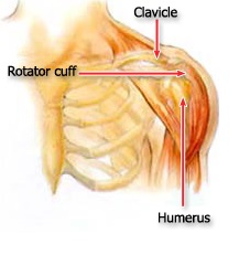
This simplified illustration of the shoulder highlights the major components of the joint.


The head of your upper arm bone fits into a rounded socket in your shoulder blade. This socket is called the glenoid. A combination of muscles and tendons keeps your arm bone centered in your shoulder socket. These tissues are called the rotator cuff. They cover the head of your upper arm bone and attach it to your shoulder blade.

Cause

Most shoulder problems fall into four major categories:

  • Tendon inflammation (bursitis or tendinitis) or tendon tear
  • Instability
  • Arthritis
  • Fracture (broken bone)

Other much less common causes of shoulder pain are tumors, infection, and nerve-related problems.

Bursitis

Bursae are small, fluid-filled sacs that are located in joints throughout the body, including the shoulder. They act as cushions between bones and the overlying soft tissues, and help reduce friction between the gliding muscles and the bone.

Sometimes, excessive use of the shoulder leads to inflammation and swelling of the bursa between the rotator cuff and part of the shoulder blade known as the acromion. The result is a condition known as subacromial bursitis. Bursitis often occurs in association with rotator cuff tendinitis. The many tissues in the shoulder can become inflamed and painful. Many daily activities, such as combing your hair or getting dressed, may become difficult.

Tendinitis

A tendon is a cord that connects muscle to bone. Most tendinitis is a result of a wearing down of the tendon that occurs slowly over time, much like the wearing process on the sole of a shoe that eventually splits from overuse.

Generally, tendinitis is one of two types:

  • Acute. Excessive ball throwing or other overhead activities during work or sport can lead to acute tendinitis.
  • Chronic. Degenerative diseases like arthritis or repetitive wear and tear due to age, can lead to chronic tendinitis.
  • The most commonly affected tendons in the shoulder are the four rotator cuff tendons and one of the biceps tendons. The rotator cuff is made up of four small muscles and their tendons that cover the head of your upper arm bone and keep it in the shoulder socket. Your rotator cuff helps provide shoulder motion and stability.

Tendon Tears

Splitting and tearing of tendons may result from acute injury or degenerative changes in the tendons due to advancing age, long-term overuse and wear and tear, or a sudden injury. These tears may be partial or may completely split the tendon into two pieces. In most cases of complete tears, the tendon is pulled away from its attachment to the bone. Rotator cuff and biceps tendon injuries are among the most common of these injuries.

Impingement

Shoulder impingement occurs when the top of the shoulder blade (acromion) puts pressure on the underlying soft tissues when the arm is lifted away from the body. As the arm is lifted, the acromion rubs, or "impinges" on, the rotator cuff tendons and bursa. This can lead to bursitis and tendinitis, causing pain and limiting movement. Over time, severe impingement can even lead to a rotator cuff tear.

Instability

Shoulder instability occurs when the head of the upper arm bone is forced out of the shoulder socket. This can happen as a result of a sudden injury or from overuse.

Shoulder dislocations can be partial, with the ball of the upper arm coming just partially out of the socket. This is called a subluxation. A complete dislocation means the ball comes all the way out of the socket.

Once the ligaments, tendons, and muscles around the shoulder become loose or torn, dislocations can occur repeatedly. Recurring dislocations, which may be partial or complete, cause pain and unsteadiness when you raise your arm or move it away from your body. Repeated episodes of subluxations or dislocations lead to an increased risk of developing arthritis in the joint.

Arthritis

Shoulder pain can also result from arthritis. There are many types of arthritis. The most common type of arthritis in the shoulder is osteoarthritis, also known as "wear and tear" arthritis. Symptoms, such as swelling, pain, and stiffness, typically begin during middle age. Osteoarthritis develops slowly and the pain it causes worsens over time.

Osteoarthritis, may be related to sports or work injuries and chronic wear and tear. Other types of arthritis can be related to rotator cuff tears, infection, or an inflammation of the joint lining.

Often people will avoid shoulder movements in an attempt to lessen arthritis pain. This sometimes leads to a tightening or stiffening of the soft tissue parts of the joint, resulting in a painful restriction of motion.

Fracture

Fractures are broken bones. Shoulder fractures commonly involve the clavicle (collarbone), humerus (upper arm bone), and scapula (shoulder blade).

Shoulder fractures in older patients are often the result of a fall from standing height. In younger patients, shoulder fractures are often caused by a high energy injury, such as a motor vehicle accident or contact sports injury.

Fractures often cause severe pain, swelling, and bruising about the shoulder.

Doctor's Examination

In the case of an acute injury causing intense pain, seek medical care as soon as possible. If the pain is less severe, it may be safe to rest a few days to see if time will resolve the problem. If symptoms persist, see a doctor.

Your doctor will conduct a thorough evaluation in order to determine the cause of your shoulder pain and provide you with treatment options.

Medical History

The first step in the evaluation is a thorough medical history. Your doctor may ask how and when the pain started, whether it has occurred before and how it was treated, and other questions to help determine both your general health and the possible causes of your shoulder problem. Because most shoulder conditions are aggravated by specific activities, and relieved by specific activities, a medical history can be a valuable tool in finding the source of your pain.

Physical Examination

A comprehensive examination will be required to find the causes of your shoulder pain. Your doctor will look for physical abnormalities, swelling, deformity or muscle weakness, and check for tender areas. He or she will observe your shoulder range of motion and strength.

Tests

Your doctor may order specific tests to help identify the cause of your pain and any other problems.

X-rays. These pictures will show any injuries to the bones that make up your shoulder joint.

Magnetic resonance imaging (MRI) and ultrasound. Thes imaging studies create better pictures of soft tissues. It may help your doctor identify injuries to the ligaments and tendons surrounding your shoulder joint.

Computed tomography (CT) scan. This tool combines x-rays with computer technology to produce a very detailed view of the bones in the shoulder area.

Electrical studies. Your doctor may order a tests, such as the EMG (electromyogram), to evaluate nerve function.

Arthrogram. During this x-ray study, dye is injected into the shoulder to better show the joint and its surrounding muscles and tendons.

Arthroscopy. In this surgical procedure, your doctor looks inside the joint with a fiber-optic camera. Arthroscopy may show soft tissue injuries that are not apparent from the physical examination, x-rays, and other tests. In addition to helping find the cause of pain, arthroscopy may be used to correct the problem.

Treatment

Activity Changes

Treatment generally involves rest, altering your activities, and physical therapy to help you improve shoulder strength and flexibility. Common sense solutions such as avoiding overexertion or overdoing activities in which you normally do not participate can help to prevent shoulder pain.

Medications

Your doctor may prescribe medication to reduce inflammation and pain. If medication is prescribed to relieve pain, it should be taken only as directed. Your doctor may also recommend injections of numbing medicines or steroids to relieve pain.

Surgery

Surgery may be required to resolve some shoulder problems; however, 90 percent of patients with shoulder pain will respond to simple treatment methods such as altering activities, rest, exercise, and medication.

Certain types of shoulder problems, such as recurring dislocations and some rotator cuff tears, may not benefit from exercise. In these cases, surgery may be recommended fairly early.

Surgery can involve arthroscopy to remove scar tissue or repair torn tissues, or traditional, open procedures for larger reconstructions or shoulder replacement.

Cervical Radiculopathy

Cervical radiculopathy, commonly called a “pinched nerve” occurs when a nerve in the neck is compressed or irritated where it branches away from the spinal cord. This may cause pain that radiates into the shoulder, as well as muscle weakness and numbness that travels down the arm and into the hand.
Cervical radiculopathy is often caused by “wear and tear” changes that occur in the spine as we age, such as arthritis. In younger people, it is most often caused by a sudden injury that results in a herniated disk.
In most cases, cervical radiculopathy responds well to conservative treatment that includes medication and physical therapy.
 

Cause

Cervical radiculopathy most often arises from degenerative changes that occur in the spine as we age or from an injury that causes a herniated, or bulging, intervertebral disk.
Degenerative changes. As the disks in the spine age, they lose height and begin to bulge. They also lose water content, begin to dry out, and become stiffer. This problem causes settling, or collapse, of the disk spaces and loss of disk space height.
As the disks lose height, the vertebrae move closer together. The body responds to the collapsed disk by forming more bone —called bone spurs—around the disk to strengthen it. These bone spurs contribute to the stiffening of the spine.
They may also narrow the foramen—the small openings on each side of the spinal column where the nerve roots exit—and pinch the nerve root.
Degenerative changes in the disks are often called arthritis or spondylosis. These changes are normal and they occur in everyone. In fact, nearly half of all people middle-aged and older have worn disks and pinched nerves that do not cause painful symptoms. It is not known why some patients develop symptoms and others do not.
Herniated disk. A disk herniates when its jelly-like center (nucleus) pushes against its outer ring (annulus). If the disk is very worn or injured, the nucleus may squeeze all the way through. When the herniated disk bulges out toward the spinal canal, it puts pressure on the sensitive nerve root, causing pain and weakness in the area the nerve supplies.
A herniated disk often occurs with lifting, pulling, bending, or twisting movements.

Symptoms

In most cases, the pain of cervical radiculopathy starts at the neck and travels down the arm in the area served by the damaged nerve. This pain is usually described as burning or sharp. Certain neck movements—like extending or straining the neck or turning the head—may increase the pain.

Other symptoms include:

  • Tingling or the feeling of “pins and needles” in the fingers or hand
  • Weakness in the muscles of the arm, shoulder, or hand
  • Loss of sensation
Some patients report that pain decreases when they place their hands on top of their head. This movement
may temporarily relieve pressure on the nerve root.
Doctor Examination
Physical Examination
After discussing your medical history and general health, your doctor will ask you about your symptoms. He or she will then examine your neck, shoulder, arms and hands—looking for muscle weakness, loss of sensation, or any change in your reflexes.
Your doctor may also ask you to perform certain neck and arm movements to try to recreate and/or relieve your symptoms.

Tests

X-rays. These provide images of dense structures, such as bone. An x-ray will show the alignment of bones along your neck. It can also reveal whether there is any narrowing of the foramen and damage to the disks.
Computed tomography (CT) scans. More detailed than a plain x-ray, a CT scan can help your doctor determine whether you have developed bone spurs near the foramen in your cervical spine.
Magnetic resonance imaging (MRI) scans. These studies create better images of the body’s soft tissues. An MRI of the neck can show if your nerve compression is caused by damage to soft tissues—such as a bulging or herniated disk. It can also help your doctor determine whether there is any damage to your spinal cord or nerve roots.
Electromyography (EMG). Electromyography measures the electrical impulses of the muscles at rest and during contractions. Nerve conduction studies are often done along with EMG to determine if a nerve is functioning normally.
Together, these tests can help your doctor determine whether your symptoms are caused by pressure on spinal nerve roots and nerve damage or by another condition that causes damage to nerves, such as diabetes.

Treatment

It is important to note that the majority of patients with cervical radiculopathy get better over time and do not need treatment. For some patients, the pain goes away relatively quickly—in days or weeks. For others, it may take longer.
It is also common for cervical radiculopathy that has improved to return at some point in the future. Even when this occurs, it usually gets better without any specific treatment.
In some cases, cervical radiculopathy does not improve, however. These patients require evaluation and treatment.

Nonsurgical Treatment

Initial treatment for cervical radiculopathy is nonsurgical. Nonsurgical treatment options include:
Soft cervical collar. This is a padded ring that wraps around the neck and is held in place with Velcro. Your doctor may advise you to wear a soft cervical collar to allow the muscles in your neck to rest and to limit neck motion. This can help decrease the pinching of the nerve roots that accompany movement of the neck. A soft collar should only be worn for a short period of time since long-term wear may decrease the strength of the muscles in your neck.
Physical therapy. Specific exercises can help relieve pain, strengthen neck muscles, and improve range of motion. In some cases, traction can be used to gently stretch the joints and muscles of the neck.
Medications. In some cases, medications can help improve your symptoms.
Nonsteroidal anti-inflammatory drugs (NSAIDs). NSAIDs, including aspirin, ibuprofen, and naproxen, may provide relief if your pain is caused by nerve irritation or inflammation.
Oral corticosteroids. A short course of oral corticosteroids may help relieve pain by reducing swelling and inflammation around the nerve.
Narcotics. These medications are reserved for patients with severe pain that is not relieved by other options. Narcotics are usually prescribed for a limited time only.

Interventional Pain Management

Cervical Epidural Corticosteroid Injections. In this procedure, a steroidal medication is injected near the affected nerve to reduce local inflammation. The injection may be placed between the laminae (midline posterior approach) or in the foramenal opening (selective nerve root injection). Steroidal injections work by promoting healing of the inflamed and injured nerve root causing the pain symproms.

Surgical Treatment

If after a period of time nonsurgical treatment does not relieve your symptoms, your doctor may recommend surgery.
There are several surgical procedures to treat cervical radiculopathy. The procedure your doctor recommends will depend
on many factors, including what symptoms you are experiencing and the location of the involved nerve root.

Source: http://orthoinfo.aaos.org/topic.cfm?topic=A00332

Cervical Disc Injury (Herniation / Bulges)

Displacement, Cervical Intervertebral Disc Without Myelopathy

 
Displacement of a cervical intervertebral disc refers to protrusion or herniation of the disc between two adjacent bones (vertebrae) of the cervical spine in the neck (vertebrae C2 through C7). Note that there is no disc between the skull and C1 or between C1 and C2. Although displacement is commonly referred to as a slipped disc, the disc does not actually slip.

The discs between each vertebra form a cushion that absorbs shock and allows movement of the neck. The discs are composed of an inner gel-like material (nucleus pulposus) and an outer ring of tough, fibrous material (annulus fibrosis). Sometimes the fibrous material develops a weak area that allows the nucleus pulposus to intrude into the spinal canal (disc displacement or herniation). Depending on the site of the intrusion, the disc may compress either the spinal cord or the exiting nerves, or both. Pressure on an exiting cervical nerve root where it exits the spinal canal can cause changes in sensory (touch, pinprick, temperature), motor (muscle strength), and reflex function in the innervated areas (upper limb). These types of changes are collectively referred to as radiculopathy; however, disc displacement may also occur without radiculopathy. Cervical radiculopathy also may be caused by tumorsinfection, or vertebral fracture. Disruption of the annulus fibrosis itself may also cause symptoms (annular disruption, distension, or tear). This can allow the nucleus pulposus to leak out of the disc, causing an intense and painful chemical inflammation (radiculitis).

Disc herniations (commonly called “soft discs” in the neck) tend to occur in younger adults who have only mild loss of disc height (disc degeneration) and thus have enough disc material still present to produce a protrusion or herniation. The same radiculopathy symptoms and signs on exam can be produced without a disc herniation in older individuals who no longer have enough disc height (disc material) to produce a herniation, but rather have arthritic spurs (osteophytes) as the structure that pinches the nerve root. In these older individuals the term used to indicate the cause of the radiculopathy is “hard disc,” meaning bone spur or “bony bar.”


Causation and Known Risk Factors There are no prospective cohort studies to assess causation of cervical disc herniation. There are case-control studies that pose the question of whether smoking, heavy lifting, and being a professional driver increase the risk of developing a cervical disc herniation.

Diagnosis

History: Important items to note in the history include: information about pain (onset, location, quantity, quality, setting, aggravating and alleviating factors, associated symptoms), percentage of pain that is axial (neck) vs. peripheral (upper limb) pain, and history of neck injury. Disc-related pain without nerve root involvement may be vague and diffuse. Radicular pain from nerve root compression typically follows a dermatomal pattern in upper limb; neck pain may be paradoxically absent. The pain may have begun with no apparent cause, or there may be a history of injury to the neck. Some episodes begin during or shortly after the person does a “low violence” activity that the individual has done many times before, and this “minor trauma” may be blamed for the event by both health care providers and patients. The location of the pain, sensory loss, and muscle weakness in the limb usually allow the physician to determine which nerve root is most likely to be compressed by a disc herniation.

These individuals sometimes rest the symptomatic upper extremity on the top of their head to decrease pain. Coughing or sneezing makes the pain worse, and affected individuals may report that they are more comfortable sleeping in a reclining chair than in a bed. If treatment is not sought, individuals may notice increasing weakness in the affected limb. A history of prior or existing systemic illness should be obtained, including chronic disease (e.g., diabetes, heart disease, atherosclerosis, nervous system disorders, arthritis, infections, malignancies, or weight loss).

Physical exam: Cervical intervertebral disc displacement usually limits range of motion of the neck. The exam may show that neck movement aggravates pain, particularly when bending the head backward (hyperextension) and turning the head from side to side (rotation). The manual application of cervical compression and distraction during the physical exam may help to differentiate between disc pain and pain from other causes. Pain may increase when downward pressure is applied to the top of the head (cervical compression test) and may be relieved by traction (cervical distraction test). Examination should include assessment of muscle strength and changes in sensation and reflexes in the upper extremities. Lower extremities may be examined to rule out signs of myelopathy.

Tests: Laboratory blood tests are usually not necessary, but may include erythrocyte sedimentation rate (ESR) to evaluate inflammation, white blood count analysis to rule out infection, rheumatoid factor, thyroid and parathyroid studies, and liver function studies. Human leukocyte antigens may be typed. Results of these tests help rule out other conditions.

Imaging studies show the extent of degenerative changes, but do not give any information about function. Plain x-rays show narrowing of the disc space and bone spur (osteophyte) formation, if present, as well as possible metastatic disease, spinal deformity, and spine stability. If mechanical instability is suspected as a cause of recurrent pain, it can be documented by x-rays taken with the neck bent forward (flexion) and bent backward (hyperextension).

MRI or myelography combined with CT are considered the best ways to diagnose a herniated cervical disc. Electromyography (EMG) may distinguish nerve root compression from a peripheral nerve problem such as carpal tunnel syndrome or ulnar nerve entrapment. Nevertheless, a normal EMG does not rule out nerve root compression. As in the lumbar spine, asymptomatic herniations are frequently seen in normal volunteers. For this reason, disc herniations on imaging studies must correlate with the clinical signs of nerve root deficit observed on physical examination.

Treatment Conservative therapy is the first line of treatment except in cases of severe or progressive neurologic compression that is usually in a specific area of skin supplied by a specific spinal nerve (dermatome) and can be matched to an MRI with disc protrusion at the same level. Bed rest is rarely indicated. Intermittent traction may be applied, and the individual may be taught to use intermittent traction at home.

Non-steroidal anti-inflammatory drugs (NSAIDs) may be given to relieve pain and decrease inflammation. Either oral or injected corticosteroids (“cortisone”) are commonly prescribed if there is severe radicular arm pain. If pain is severe, a narcotic may be added; in some cases, an antidepressant or an anticonvulsant may be used for its analgesic effect. If anxiety and tension are prominent, sedatives may be helpful. Muscle relaxants are frequently prescribed; however, their effectiveness probably is due to their sedative action. Narcotics, sedatives, and muscle relaxants are ideally used only for brief periods. Ongoing use should be weighed against the potential for addiction or abuse. Other treatments such as ice, heat, massage, and ultrasound therapy may help relieve pain.

As symptoms subside, activity is gradually increased and includes physical therapy to strengthen and mobilize the muscles of the neck and shoulder. An independent home exercise program is an essential component of any physical therapy. Good posture and frequent changes in position may help prevent fatigue and decrease pain. Preventive and maintenance measures, such as exercise, stress management, and proper body mechanics, should be continued indefinitely. If there is no improvement during the first 2 weeks, or if pain is still disabling after 6 weeks, further evaluation is necessary.

Most cases of cervical disc displacement with or without radiculopathy can be managed conservatively.

Pain Interventional procedures are within the scope of conservative treatments for disc related pain withor without Radiculopathy( neck pain radiating to the arm with numbness and/or tingling sensations in the arm). These include mainly Cervical Epidural Injections done under Flourospcopic Guidance (live X-ray) where medication (corticosteroid) is delivered directly to the area of the inflamed disc or nerve noot.

However, surgery is indicated in cases where (1) pain management has failed, and the individual has intractable upper limb pain with imaging evidence of a correlating nerve root compression; (2) there is mechanical instability of the spine associated with disc herniation; (3) signs of neurological deficits are increasing (e.g., progressive or severe muscle weakness or severe arm pain with objective signs of nerve root compression on imaging); or (4) the disc herniation is massive and compresses the spinal cord, causing bowel and/or bladder control impairment, lower extremity weakness, sensory loss, or gait disturbance.

Surgery involves removal of the protruding nucleus pulposus (discectomy). The traditional method for removal of the disc is open discectomy under general anesthesia. The discectomy is most often done through an anterior approach (incision in the front of the neck), and is most often accompanied by a simultaneous fusion, frequently supplemented by use of a plate and screws. An alternative that is occasionally chosen is posterior (back of the neck incision) discectomy in which a portion of the vertebra that acts as a roof (lamina) over the spinal nerve is removed, creating a small window into the spine. The surgeon then removes the herniated disc material through this opening.

An alternative for younger patients who do not have facet arthritis or other significant aging change is anterior discectomy with simultaneous artificial disc replacement. This procedure appears to give early results as good as anterior cervical discectomy and fusion, but long term results are not yet known or published (Gebremariam).

Rehabilitation The primary focus of rehabilitation for a cervical intervertebral disc displacement without myelopathy is to decrease symptoms and increase function. Although exercise may be uncomfortable initially, individuals must be instructed in the benefits of ongoing exercise in managing the symptoms.

The first goal is to decrease symptoms, primarily pain. In combination with pharmacological management, modalities such as heat and cold can be used. Immobilization with a soft collar is rarely indicated; however with significant soft tissue pain, it might be necessary for a very short period of time (up to 3 days). While managing pain, individuals can be instructed in gentle exercises (Boyce). Due to the variability in response, the treating practitioner must pay careful attention to tolerance to treatment. Initial exercises may include isometrics, stretching and/or gentle range of motion. Spinal manual therapy may reduce symptoms when combined with active treatment. Postural training should be initiated as soon as tolerated by the individual.

Once symptoms subside and range of motion is restored, the individual should progress to strengthening and stabilization exercises of the neck, shoulders and upper trunk (Ylinen). Limited treatment with cervical traction has been shown to be beneficial for neck pain when done in conjunction with exercises, although traction must be carefully administered to avoid adverse response.

The individual should also be instructed in a home exercise program to complement the supervised rehabilitation, and trained to care for and protect the neck from recurrence of symptoms. An ergonomic evaluation can prove helpful in avoiding or modifying activities and work positions that may aggravate the symptoms. Psychotherapy may be indicated to support the individual and identify associated factors that may contribute to the symptoms. A short course of cognitive pain management may be beneficial for individuals experiencing psychological distress or lack of improvement with treatment (Klaber Moffett).
Complications Worsening of the condition (enlargement or increasing size of the disc herniation) may cause pressure on the spinal cord as well as on additional nerve roots. Functional disturbances and/or pathological changes in the spinal cord (myelopathy) may occur as a result of the displaced disc pressing on the spinal cord. Muscular atrophy and sensory disorders may occur as the result of nerve root compression.

Source: http://www.mdguidelines.com/displacement-cervical-intervertebral-disc-without-myelopathy

Facet Joint Arthritis

INTRODUCTION

Arthritis of the spinal facet joints can be a source of significant neck and low back pain. Aligned on the back of the spinal column, the facet joints link each vertebra together. Articular cartilage covers the surfaces where these joints meet. Like other joints in the body that are covered with articular cartilage, the lumbar facet joints can be affected by arthritis.

ANATOMY:

The human spine is made up of 24 spinal bones, called vertebrae. Vertebrae are stacked on top of one another to create the spinal column. The spinal column gives the body its form. It is the body's main upright support.
The back portion of the spinal column forms a bony ring. When the vertebrae are stacked on top of each other, these bony rings create a hollow tube. This tube, called the spinal canal, surrounds the spinal cord as it passes through the spine. Just as the skull protects the brain, the bones of the spinal column protect the spinal cord.
Between the vertebrae of each spinal segment are two facet joints. The facet joints are located on the back of the spinal column. There are two facet joints between each pair of vertebrae, one on each side of the spine. A facet joint is made of small, bony knobs that line up along the back of the spine. Where these knobs meet, they form a joint that connects the two vertebrae. The alignment of the facet joints of the lumbar spine allows freedom of movement as you bend forward and back. The surfaces of the facet joints are covered by articular cartilage. Articular cartilage is a smooth, rubbery material that covers the ends of most joints. It allows the bone ends to move against each other smoothly, without friction.

CAUSES

Why do I have this problem of neck and back pain?
Normally, the facet joints fit together snugly and glide smoothly, without pressure. If pressure builds where the joint meets, the cartilage on the joint surfaces wears off, or erodes.
Each segment in the spine has three main points of movement, the intervertebral disc and the two facet joints. Injury or problems in any one of these structures affects the other two. As a disc thins with aging and from daily wear and tear, the space between two spinal vertebrae shrinks. This causes the facet joints to press together. Wear and tear of the facet joints leads to the pain of arthritis casuing back pain.
The body responds to this extra pressure by developing bone spurs. As the spurs form around the edges of the facet joints, the joints become enlarged. This is called hypertrophy. Eventually, the joint surfaces become arthritic. When the articular cartilage degenerates, or wears away, the bone underneath is uncovered and rubs against bone. The joint becomes inflamed, swollen, and painful.
Facet joint arthritis develops slowly over a long period of time. This is partly because spinal degeneration in later life is the main cause of facet joint arthritis. Symptoms rarely develop immediately when degeneration is causing the problems.
However, rapid movements, heavy twisting, or backward motions in the spine can injure a facet joint, leading to immediate symptoms.
Facet joints can also become arthritic due to a neck or back injury earlier in life. Fractures, torn ligaments, and disc problems can all cause abnormal movement and alignment, putting extra stress on the surfaces of the facet joints.

SYMPTOMS

What does the condition feel like?
Pain from facet joint arthritis is usually worse after resting or sleeping. Also, bending the neck or trunk sideways or backward usually produces pain on the same side as the arthritic facet joint. For example, if you lie on your stomach on a flat surface and raise your upper body, you hyperextend the lumbar spine. Similarly if you tilt your head upwards, you hyperextend ghe cervical spine. This increases pressure on the facet joints and can cause pain if there is facet joint arthritis.
Image result for cervical facet arthritis
cervical facet pain
Image result for lumbar facet pain pattern
lumbar facet pain pattern
thoracic facet pain pattern
Pain may be felt in the center of the low back and can spread into one or both buttocks. Sometimes the pain spreads into the thighs, but it rarely goes below the knee. Numbness and tingling, the symptoms of nerve compression, are usually not felt because facet arthritis generally causes only mechanical pain. Mechanical pain comes from abnormal movement in the spine. For the cervical area, pain is localized the back of your neck and can spread to the shoulder blades or can cause headaches. For the thoracic or midback area, pain is localized at the level of joint involvement.
However, symptoms of nerve compression can sometimes occur at the same time as the facet joint pain. The arthritis can cause bone spurs at the edges of the facet joint. These bone spurs may form in the opening where the nerve root leaves the spinal canal. This opening is called the neural foramen. If the bone spurs rub against the nerve root, the nerve can become inflamed and irritated. This nerve irritation can cause symptoms where the nerve travels. These symptoms may include numbness, tingling, slowed reflexes, and muscle weakness in either the legs or the arms.

DIAGNOSIS

How do doctors diagnose the problem?
Diagnosis begins with a complete history and physical examination. Your doctor will ask questions about your symptoms and how your problem is affecting your daily activities. This will include questions about where you feel pain and if you have numbness or weakness in your legs or arms. Your doctor will also want to know what positions or activities make your symptoms worse or better.
Then the doctor performs a physical exam to determine which neck or back movements cause pain or other symptoms.
Your skin sensation, muscle strength, and reflexes are also tested.
X-rays can show if there are problems in the bone tissue in and near the facet joints. The images can show if degeneration has caused the space between the vertebrae to collapse and may show if bone spurs have developed near the facet joints.
When more information is needed, your doctor may order magnetic resonance imaging (MRI). The MRI machine uses magnetic waves rather than X-rays to show the soft tissues of the body. This test gives a clear picture of the facet joints to see whether they are enlarged or swollen. This machine creates pictures that look like slices of the area your doctor is interested in. The test does not require dye or a needle.
A computed tomography (CT) scan may be ordered. This is a detailed X-ray that lets your doctor see slices of bone tissue. The image can show whether the surface of the joint has eroded and whether bone spurs have developed.
A diagnostic injection done by a Physician specializing in Pain Management may be used to locate the source of pain.
The doctor uses a special needle to inject a local anesthetic (numbing medication) into either the joint or into the nerve that goes to the joint. The doctor watches the needle on a fluoroscope (live X-Ray) to make sure it reaches the correct spot. A fluoroscope is a special X-ray television that allows the doctor to see your spine and the needle as it moves. Once the doctor is sure the needle is in the right place, the medicine is injected. The results from the injection help the doctor make the diagnosis. If pain goes away, even if temporarily for a few hours, it helps confirm the source of pain.

TREATMENT OPTIONS

What treatment options are available?

NONSURGICAL TREATMENT:

BED REST: During acute pain flare-ups, facet joint arthritis is mainly treated nonsurgically. At first, doctors may prescribe a short period of rest, one to two days at most, to calm inflammation and pain. Patients may find added relief by curling up to sleep on a firm mattress or by lying back with their knees bent and supported. These positions take pressure off the facet joints.
MEDICATIONS: Your doctor may prescribe anti-inflammatory medication, such as a nonsteroidal anti-inflammatory drug (NSAID) or aspirin. Muscle relaxants are occasionally used to calm muscles that are in spasm. Oral steroid medicine in tapering dosages may also be prescribed for painflare-ups.
PHYSICAL THERAPY: Patients often work with a Physical Therapist. By evaluating a patient's condition, the therapist can assign positions and exercises to ease symptoms. The therapist may recommend traction. Traction is a common treatment for this condition. It gently stretches the neck orlow back and takes pressure off the facet joints. The therapist may also prescribe strengthening and aerobic exercises. Strengthening exercises focus on improving the strength and control of the spinal and abdominal muscles. Aerobic exercises are used to improve heart and lung health and increase endurance in the spinal muscles. Stationary biking offers a good aerobic treatment and keeps the spine bent slightly forward, a position that gives relief to many patients with lumbar facet joint arthritis.
CHIROPRACTIC: Spinal manipulation done by a Chiropractor can sometimes provide short-term relief of pain from facet arthritis. Commonly thought of as an adjustment, spinal manipulation stretches the tissues surrounding the facet joint and helps reset the sensitivity of the spinal nerves and muscles. It involves a high-impulse stretch of the spinal joints and is characterized by the sound of popping as the stretch is done. However, it doesn't seem to provide effective long-term help when used routinely for chronic conditions.

INTERVENTIONAL PAIN MANAGEMENT:

FACET JOINT INJECTIONS:
Patients who still have pain after trying various treatments may require injections into the facet joint; this is done by a Pain Management Specialist. The procedure to inject the medication into the joint itself is similar to the diagnostic injection described earlier. A steroid medication is occasionally used instead of the anesthetic. Steriodal injection directly into the facet joints involved give long-term pain relief that may last months to years. Doctors often have their patients resume physical therapy treatments following an injection.

RADIOFREQUENCY ABLATION:

If previous facet joint injections / blocks give releif that is significant but temporary, Radiofreqency Ablation can be done as a treatment option to inactivate the small tiny nerves that provide sensation to the facet joints involved causing pain.
This is done by using special needles positioned where those nerves are located (called the medial branch nerves to the facet joints) under flouroscopic (live X-ray) guidance. Radiofrequency energy is used to heat up the tip of these special needles to creat a lesion that inactivates / cauterizes the target nerves. Releif of pain after the procedure follows in the following weeks over a gradual onset and the pain relief provided is long term, lasting for months to years. This procedure can be repeated again if the pain recurs.

SURGERY:

People with facet joint arthritis rarely need surgery. However, facet joint arthritis is a primary source of chronic neck and low back pain about 70 percent of the time. After trying other types of treatment, some of these patients may eventually require surgery. There are several types of surgery for facet joint arthritis. The two primary operations are:
FACET RHIZOTOMY: Rhizotomy describes a surgical procedure in which a nerve is purposely cut or destroyed. Facet rhizotomy involves severing one of the small nerves that goes to the facet joint. The intent of the procedure is to stop the transmission of pain impulses along this nerve. The nerve is identified using a diagnostic injection (described earlier).
Then the surgeon inserts a large, hollow needle through the tissues in the low back. A special probe is inserted through the needle, and a fluoroscope is used to guide the probe toward the nerve. The probe is slowly heated until the nerve is severed.
POSTERIOR CERVICAL OR LUMBAR FUSION: Facet joint arthritis mainly causes mechanical pain, the type of pain caused by wear and tear in the parts of the spine. Posterior cervical or lumbar fusion for facet joint arthritis is mainly used to stop movement of the painful joints by joining two or more vertebrae into one solid bone (fusion). This keeps the bones and painful facet joints from moving. In this procedure, the surgeon lays small grafts of bone over the back of the spine.
Most surgeons will also apply metal plates and screws to prevent the two vertebrae from moving. This protects the graft so it can heal better and faster.

Mononeuritis multiplex

Mononeuritis multiplex

Mononeuritis multiplex is a nervous system disorder that involves damage to at least two separate nerve areas.

Causes

Mononeuritis multiplex is a form of damage to one or more peripheral nerves — the nerves outside the brain and spinal cord. It is a group of symptoms (syndrome), not a disease.

However, certain diseases can cause the injury or nerve damage that leads to the symptoms of mononeuritis multiplex.

Common conditions include:

  • Blood vessel diseases such as polyarteritis nodosa
  • Connective tissue diseases such as rheumatoid arthritis or systemic lupus erythematosus (the most common cause in children)
  • Diabetes mellitus

Less common causes include:

  • Amyloidosis
  • Blood disorders (such as hypereosinophilia and cryoglobulinemia)
  • Infections such as Lyme disease
  • Leprosy
  • Sarcoidosis
  • Sjogren syndrome
  • Wegener’s granulomatosis

Symptoms

Symptoms will depend on the specific nerves involved, and may include:

  • Loss of bladder or bowel control
  • Loss of sensation in one or more areas of the body
  • Paralysis in one or more areas of the body
  • Tingling, burning, pain, or other abnormal sensations in one or more areas of the body
  • Weakness in one or more areas of the body

Exams and Tests

A detailed history is needed to determine the possible cause of the disorder. Examination and neuromuscular testing may show a loss of feeling and movement due to problems with specific nerves. Reflexes may be abnormal.

To diagnose mononeuritis multiplex, there usually needs to be problems with two or more unrelated nerve areas. Common nerves affected are the:

  • Axillary nerve in either arm and shoulder
  • Common peroneal nerve in the lower leg
  • Distal median nerve to the hand
  • Femoral nerve in the thigh
  • Radial nerve in the arm
  • Sciatic nerve in the back of the leg
  • Ulnar nerve in the arm

Tests may include:

  • Electromyogram (EMG, a recording of electrical activity in the muscles)
  • Nerve biopsy to examine the nerve under a microscope
  • Nerve conduction tests to measure how fast nerve impulses move along the nerve

Other tests may include:

  • Antinuclear antibody panel (ANA)
  • Blood chemistry tests
  • C-reactive protein
  • Imaging scans
  • Pregnancy test
  • Rheumatoid factor
  • Sedimentation rate
  • Thyroid tests
  • X-rays

Treatment

The goals of treatment are to:

  • Treat the illness that is causing the problem, if possible
  • Provide supportive care to maximize independence
  • Control symptoms (this may include controlling blood sugar levels for diabetics, nutritional supplementation, or medically treating conditions)

To improve independence, treatments may include:

  • Occupational therapy
  • Orthopedic help (for example, appliances such as wheelchairs, braces, and splints)
  • Physical therapy (for example, exercises and retraining to increase muscle strength)
  • Vocational therapy

Safety is an important consideration for people with sensation or movement difficulties. Lack of muscle control and decreased sensation may increase the risk of falls or injuries. Safety measures for people with movement difficulty include:

  • Adequate lighting (including leaving lights on at night)
  • Railings
  • Removing obstacles (such as loose rugs that may slip on the floor)
  • Testing water temperature before bathing
  • Wearing protective shoes (no open toes or high heels)

Check shoes often for grit or rough spots that may injure the feet.

People with decreased sensation should check their feet (or other affected area) often for bruises, open skin areas, or other injuries that may go unnoticed. These injuries may become severely infected because the pain nerves of the area are not signaling the injury.

People with mononeuropathy multiplex are prone to new nerve injuries at pressure points such as knees and elbows. They should avoid putting pressure on these areas, for example by not leaning on the elbows, crossing the knees, or holding similar positions.

Medications that may help include:

  • Over-the-counter pain medicines (ibuprofen or acetaminophen) or prescription pain medications may be needed to control pain (neuralgia).
  • Anticonvulsants (gabapentin, phenytoin, carbamazepine, or pregabalin) or antidepressants (amitriptyline, nortriptyline, or duloxetine), may be used to reduce stabbing pains.

Whenever possible, avoid or minimize the use of medications to reduce the risk of side effects.

Positioning (the use of frames to keep bedclothes off of a tender body part) and other measures may help control pain. Autonomic symptoms can be difficult to treat or respond poorly to treatment.

Outlook (Prognosis)

A full recovery is possible if the cause is found and treated, especially if the nerve damage is limited. Some people have no type of disability. Others have a partial or complete loss of movement, function, or sensation.

Nerve pain may be quite uncomfortable and can last for a long time. If this occurs, see a pain specialist to discuss all pain treatment options available to you.

Possible Complications

  • Deformity, loss of tissue or muscle mass
  • Disturbances of organ functions
  • Medication side effects
  • Repeated or unnoticed injury to the affected area due to lack of sensation
  • Relationship problems due to impotence

When to Contact a Medical Professional

Call your health care provider if you notice signs of mononeuritis multiplex.

Prevention

Preventive measures vary depending on the specific disorder. Eating a proper diet and taking medication for diabetes may help prevent mononeuritis multiplex from developing.